Evidence deep dives for Aspiration Pneumonia
Pair mechanism-level evidence with practical protocol context before discussing next steps with your veterinarian.
When Food Goes Down the Wrong Way — and the Lungs Pay the Price
One coughing fit after a regurgitation episode. A wet, rattling breath that was not there an hour ago. For dogs with conditions like megaesophagus or brachycephalic syndrome, these moments are not just scary — they can be the start of a life-threatening lung infection.
Aspiration pneumonia develops when food, water, saliva, or vomit enters the lower airways. The damage unfolds in two waves. First, gastric acid and food particles burn the delicate airway lining (chemical pneumonitis). Then, within 24-48 hours, bacteria from the mouth and gut colonize the damaged tissue and produce a full-blown infection.
The right lung takes the worst hit. The right main bronchus is wider and more vertically angled than the left, creating a gravitational funnel for aspirated material. That is why right middle and cranial lung lobe consolidation is the radiographic signature of this disease.
Why Aspiration Pneumonia Shortens Lives
Mortality rates range from 20% to 50%, depending on how quickly treatment begins and what caused the aspiration in the first place. Dogs that survive one episode remain at elevated risk for the next unless the underlying problem is controlled.
The speed of deterioration makes this condition particularly dangerous. A dog that seems mildly uncomfortable after a choking episode can progress to respiratory failure within hours as bacterial infection sets in.
For dogs living with chronic predisposing conditions — megaesophagus, brachycephalic anatomy, laryngeal paralysis — aspiration pneumonia is often what ultimately ends their life. That makes prevention the single most important longevity strategy for these dogs.
Breeds That Face the Highest Risk
Anatomy and physiology determine vulnerability:
- English Bulldog: brachycephalic airway obstruction combined with high rates of hiatal hernia and gastroesophageal reflux
- Pug: brachycephalic airway syndrome raises aspiration risk during feeding and sleep
- Labrador Retriever: prone to laryngeal paralysis and rapid, gulping eating behavior
- Irish Setter: predisposed to megaesophagus
French Bulldogs, Golden Retrievers (myasthenia gravis risk), and Great Danes (megaesophagus predisposition) also carry elevated risk. Any senior dog with laryngeal paralysis, swallowing dysfunction, or neurological disease affecting the throat muscles is a candidate.
Recognizing the Warning Signs
Within Hours of Aspiration
- Sudden wet, productive cough
- Faster breathing with visible effort
- Gagging or retching after a regurgitation or vomiting episode
- Nasal discharge that looks food-tinged
- Restlessness or refusal to lie down
- Mild fever
As Infection Takes Hold (12-48 Hours)
- Worsening respiratory distress: open-mouth breathing, neck extended forward, abdominal effort
- High fever (103-106F)
- Lethargy, weakness, no interest in food
- Blue-tinged gums and tongue (cyanosis) in severe cases
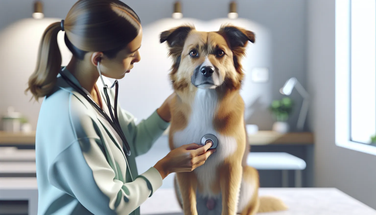
- Crackles or wheezes audible through a stethoscope
- Elevated heart rate
When It Becomes Critical
- Respiratory failure requiring supplemental oxygen
- Shock: pale gums, rapid weak pulse, cold extremities
- Acute respiratory distress syndrome (ARDS)
- Sepsis spreading from the lungs
- Collapse
What Drives Aspiration Risk
The Conditions That Set the Stage
Megaesophagus is the most common underlying cause. Chronic passive regurgitation gives aspirated material repeated chances to reach the lungs. Whether the megaesophagus stems from myasthenia gravis, idiopathic causes, or a vascular ring anomaly, the aspiration risk stays elevated for life.
Brachycephalic airway syndrome disrupts normal airway protective reflexes. Stenotic nares, elongated soft palate, and everted laryngeal saccules all interfere with the throat’s ability to seal off the trachea during swallowing.
Laryngeal paralysis prevents the larynx from opening and closing properly, allowing food and liquid to slip past during swallowing. This is especially common in older large-breed dogs, particularly Labs.
Chronic vomiting from any cause — pancreatitis, inflammatory bowel disease, gastrointestinal obstruction — raises the risk, especially in debilitated or neurologically impaired animals.
Situations That Create Danger
Anesthesia and sedation suppress the gag reflex. Proper fasting protocols (8-12 hours for food, 2-4 hours for water) and careful recovery monitoring are non-negotiable preventive measures.
Force-feeding or syringe-feeding can overwhelm swallowing reflexes when liquids are administered too rapidly or against a resisting animal.
Neurological disease — brain tumors, stroke, degenerative myelopathy — impairs the coordinated swallowing sequence.
Cleft palate creates a direct connection between the oral and nasal cavities, frequently causing aspiration pneumonia in neonatal puppies.
How Veterinarians Confirm the Diagnosis
Chest Radiographs
X-rays are the primary diagnostic tool. Classic findings include alveolar consolidation concentrated in the right middle and cranial lung lobes. One important caveat: radiographic changes can lag 12-24 hours behind clinical signs. If the first set of films looks normal but your vet still suspects aspiration, repeat imaging the next day often reveals what the first set missed.
Oxygen Monitoring
Pulse oximetry tracks oxygen saturation in real time. Arterial blood gas analysis measures the degree of hypoxemia more precisely and guides oxygen supplementation decisions. An SpO2 below 90% or PaO2 below 60 mmHg signals significant respiratory compromise.
Airway Sampling
In dogs stable enough for the procedure, tracheal wash or bronchoalveolar lavage collects samples for cytology and bacterial culture. Cytology typically reveals suppurative inflammation packed with neutrophils engulfing bacteria. Culture results shape targeted antibiotic selection.
Bloodwork
- Complete blood count: usually shows elevated neutrophils with band cells, signaling active bacterial infection
- Chemistry panel: checks for concurrent organ dysfunction
- Blood gas analysis: evaluates how well the lungs are exchanging oxygen and carbon dioxide
Preventing the Next Episode
If your dog lives with a condition that predisposes to aspiration, prevention is the most powerful tool you have.
Feeding Strategies for At-Risk Dogs
- Elevated feeding (Bailey chair or raised bowls) for dogs with megaesophagus
- Small, frequent meals instead of large ones
- Test different food consistencies — gruel, meatballs, or kibble — because individual dogs aspirate more with certain textures
- Keep your dog upright for 15-30 minutes after eating
- Supervise every meal
Managing Brachycephalic Risk
- Corrective surgery for significant airway obstruction (stenotic nares widening, soft palate resection) reduces aspiration risk
- Slow-feeder bowls prevent gulping
- No exercise immediately after meals
- Watch for signs of worsening brachycephalic syndrome
Before and After Anesthesia
- Follow fasting instructions precisely
- Rapid-sequence intubation for high-risk patients
- Keep the endotracheal tube cuff inflated until the swallowing reflex returns
- Position recovery patients with the head slightly elevated
- Close monitoring during the entire post-anesthetic window
General Prevention
- Treat the underlying condition — myasthenia gravis, laryngeal paralysis — that creates the aspiration risk
- Never syringe-feed liquids too fast
- Address chronic vomiting conditions promptly
- Weight management reduces respiratory burden in brachycephalic breeds
How Treatment Works
Oxygen First
Supplemental oxygen is the immediate priority. Delivery methods include nasal cannulas, an oxygen cage, or flow-by oxygen. Dogs in severe distress may need mechanical ventilation — a high-stakes intervention with a guarded prognosis, but one that can save lives.
IV Fluids
Fluid resuscitation supports the cardiovascular system, maintains organ perfusion, and helps the airways clear infected material. Fluid rates require careful calibration: too much fluid worsens pulmonary edema.
Antibiotics
Broad-spectrum antibiotics covering both aerobic and anaerobic bacteria start immediately while culture results are pending. Common empiric choices include:
- Ampicillin-sulbactam IV for hospitalized patients
- Amoxicillin-clavulanate for stable outpatient cases
- Fluoroquinolone plus clindamycin or metronidazole for polymicrobial coverage
Antibiotics typically continue for 3-6 weeks, extending at least 1-2 weeks past radiographic resolution.
Nebulization and Coupage
Nebulization with sterile saline hydrates airway secretions, making them easier to clear. Coupage — gentle percussive tapping on the chest wall — helps mobilize secretions from the lower airways. Hospitalized patients receive both treatments multiple times daily.
Managing the Underlying Trigger
For dogs whose aspiration was driven by vomiting or reflux:
- Maropitant (Cerenia) controls nausea
- Omeprazole or famotidine reduces gastric acid
- Metoclopramide promotes gastric motility (contraindicated if GI obstruction is present)
Nutrition During Recovery
No supplement prevents or treats aspiration pneumonia. Nutritional management during recovery is about feeding safely:
- Nothing by mouth during acute respiratory distress until the dog stabilizes
- Resume feeding with small amounts of the right-consistency food once breathing improves
- Elevated feeding position during and after every meal
- Dogs unable to eat safely by mouth may need a nasogastric or esophagostomy tube
- Adequate caloric intake supports immune function and tissue healing
For recovery-phase feeding strategy, see Post-Illness Recovery Nutrition and Immune Support Nutrition Protocol. Dogs with underlying swallowing disorders may benefit from structured feeding protocols discussed in Feeding Guide for Adult Dogs.
When Your Dog Needs a Vet
Routine monitoring is appropriate for:
- Dogs with known aspiration risk factors (megaesophagus, brachycephalic syndrome) during stable periods
- Follow-up radiographs to confirm resolution after treatment
Prompt evaluation is needed for:
- New cough after a regurgitation or vomiting episode
- Mild increase in breathing rate without distress
- Reduced appetite with mild lethargy following a choking episode
- Nasal discharge that appears food-tinged
Emergency evaluation — do not wait:
- Any acute respiratory distress (increased effort, extended neck, open-mouth breathing)
- Cyanosis (blue or gray gums)
- Fever with respiratory signs
- Collapse or severe weakness
- You witnessed food or liquid going “down the wrong way” followed by persistent coughing
Related Condition Pathways
Related Breed Longevity Guides
Related Science
- Senior Dog Screening Protocol
- Weight Management Protocol for Dogs
- Canine Frailty Signals and Early Intervention
Diet and Supplement Considerations
Aspiration pneumonia treatment is primarily medical, but nutritional support during and after recovery influences healing speed and immune resilience.
- Post-Illness Recovery Nutrition: caloric and protein targets for dogs rebuilding strength after acute respiratory illness and hospitalization.
- Immune Support Nutrition Protocol: evidence-based nutritional strategies that support immune function during infection recovery.
- Feeding Guide for Senior Dogs: senior dogs with aspiration risk factors benefit from adjusted feeding strategies that account for both nutritional needs and swallowing safety.
Verify any changes to this protocol with your veterinarian. Even seemingly minor dose or timing shifts can affect treatment outcomes.
Frequently Asked Questions
Can a dog survive aspiration pneumonia? Yes. With prompt treatment — oxygen support, IV antibiotics, and aggressive supportive care — survival rates range from 50-80%. The key factors are how quickly treatment begins, how severely the lungs are compromised when the dog arrives at the clinic, and whether the underlying cause can be controlled going forward.
How long does recovery take? Hospital stays typically last 2-7 days for moderate cases. Antibiotics continue for 3-6 weeks total. Radiographic clearing may take 2-4 weeks after clinical improvement. Most survivors return to baseline respiratory function, but full recovery can take 4-8 weeks.
Can aspiration pneumonia be prevented in dogs with megaesophagus? Elevated feeding and keeping the dog upright after meals significantly reduce the risk, but they cannot eliminate it entirely. Dogs with megaesophagus face lifelong vulnerability. The best food consistency varies by individual — some dogs do better with gruel, others with formed meatballs — and finding the right approach takes careful observation with veterinary guidance.
Is aspiration pneumonia contagious? No. The bacteria involved are normal residents of the mouth and gastrointestinal tract, not pathogens that spread between dogs. The problem is their location, not their identity — these bacteria cause harm only when they reach the lungs.
Why are brachycephalic dogs at higher risk? Their anatomy works against them on multiple fronts. Elongated soft palates, narrowed airways, and frequent gastrointestinal issues (hiatal hernia, gastroesophageal reflux) all disrupt the throat’s ability to keep food and liquid out of the trachea. These structural features raise aspiration risk during eating, drinking, and even sleep.
Medical Disclaimer
This content is for educational purposes only and does not constitute veterinary medical advice. Aspiration pneumonia is a life-threatening emergency. If your dog shows signs of respiratory distress after regurgitation, vomiting, or a choking episode, seek emergency veterinary care immediately.
References
[1] Kogan DA, Johnson LR, Sturges BK, et al. “Etiology and clinical outcome in dogs with aspiration pneumonia: 88 cases (2004-2006).” J Am Vet Med Assoc. 2008;233(11):1748-1755. [2] Tart KM, Babski DM, Lee JA. “Potential risks, prognostic indicators, and diagnostic and treatment modalities affecting survival in dogs with presumptive aspiration pneumonia: 125 cases (2005-2008).” J Vet Emerg Crit Care. 2010;20(3):319-329. [3] Proulx A, Hume DZ, Drobatz KJ, Reineke EL. “In vitro bacterial isolate susceptibility versus clinical response to antimicrobial therapy in dogs with bacterial pneumonia: 79 cases (2006-2011).” J Vet Emerg Crit Care. 2014;24(2):219-229. [4] MacPhail C. “Laryngeal disease in dogs and cats.” Vet Clin North Am Small Anim Pract. 2014;44(1):19-31. [5] Dear JD. “Bacterial pneumonia in dogs and cats: an update.” Vet Clin North Am Small Anim Pract. 2020;50(2):447-459. [6] Meola SD. “Brachycephalic airway syndrome.” Top Companion Anim Med. 2013;28(3):91-96.
Related reads
Related Reading
Continue exploring