Evidence deep dives for Corneal Ulcer
Pair mechanism-level evidence with practical protocol context before discussing next steps with your veterinarian.
A Painful Eye That Cannot Wait
A corneal ulcer is a defect in the corneal surface — and potentially deeper layers — of the eye. Ulcers range from superficial (epithelium only) to deep stromal defects that risk perforation. The depth determines urgency, treatment intensity, and whether vision can be saved.
Trauma (a scratch from vegetation, a cat claw), foreign bodies, dry eye (keratoconjunctivitis sicca), eyelid abnormalities (entropion, ectropion, distichiasis), and infection all cause corneal ulcers. In older dogs, spontaneous chronic corneal epithelial defects (SCCED, also called indolent ulcers or boxer ulcers) develop when the epithelium fails to adhere normally to underlying stroma.
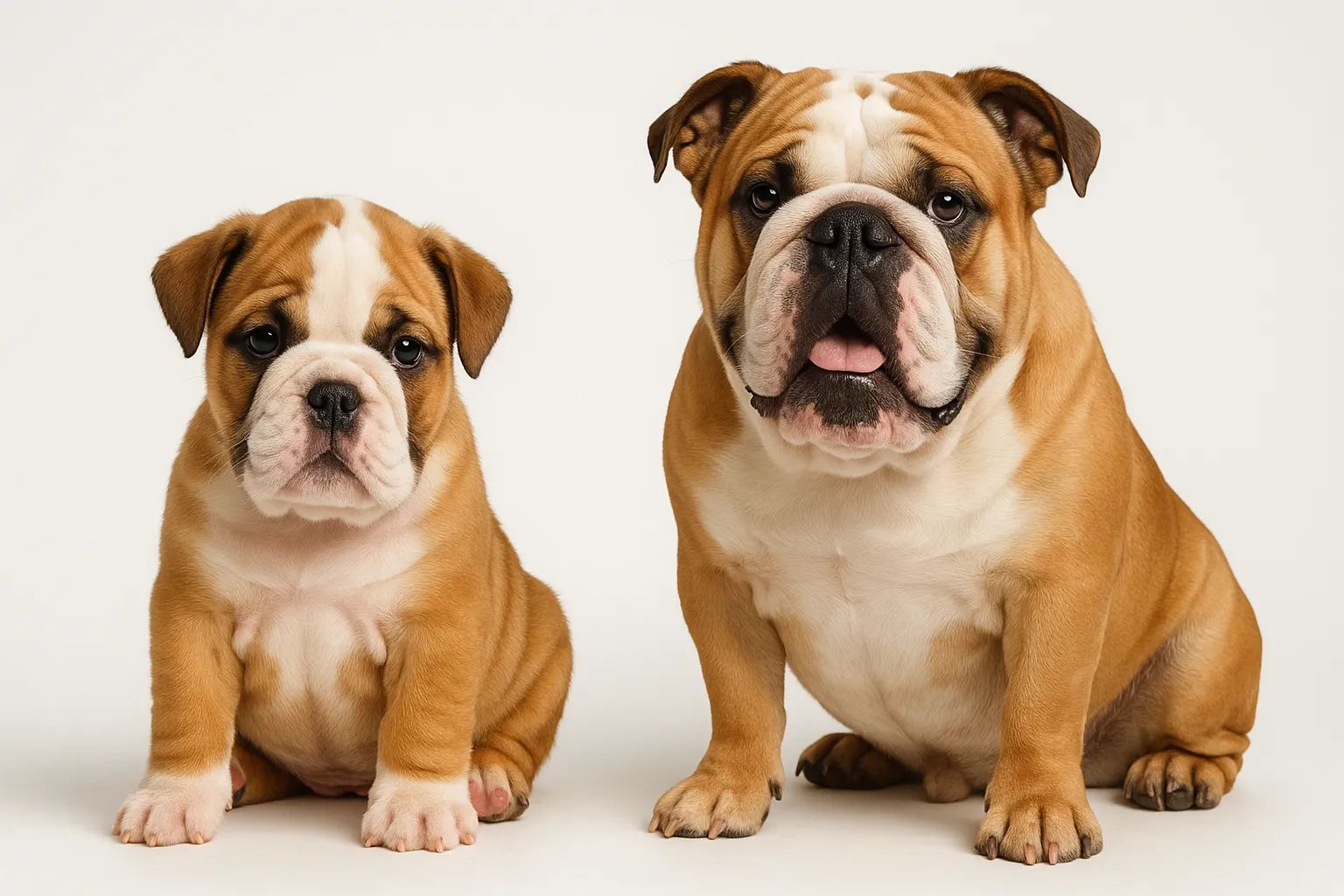
Brachycephalic breeds face dramatically elevated risk. French Bulldogs, Pugs, English Bulldogs, Shih Tzus, and Pekingese have bulging eyes that are more exposed and more vulnerable to trauma, eyelids that may not fully close, and reduced corneal sensation. These anatomical factors make corneal injury both more likely and more dangerous.
The Longevity Impact
An untreated corneal ulcer can perforate the eye within 24-72 hours in deep cases with bacterial involvement. Perforation causes loss of aqueous humor, iris prolapse, and typically permanent vision loss. That timeline makes deep or infected corneal ulcers genuine emergencies.
For brachycephalic breeds especially, corneal disease represents a recurrent, breed-defining health challenge. Repeated ulceration events cause pain, demand intensive treatment, and can produce corneal scarring that permanently reduces vision. Proactively managing underlying brachycephalic ocular abnormalities — correcting entropion surgically, performing medial canthoplasty to reduce exposure — lowers the lifetime corneal disease burden and protects quality of life.
Recognizing a Corneal Ulcer
Corneal ulcers are typically acutely painful. Dogs show characteristic discomfort signs:
- Squinting (blepharospasm) — often the most prominent and consistent sign
- Excessive tearing or eye discharge
- Pawing at the eye or rubbing the face on carpet or furniture
- Visible cloudiness or haziness on the corneal surface
- Redness of the conjunctiva (the white of the eye looks red)
- Photophobia — the dog avoids bright light and keeps the affected eye partially closed
Any dog with acute eye squinting should be seen the same day. Corneal ulcers that appear minor can worsen rapidly, particularly in brachycephalic breeds where reduced corneal sensation blunts early pain signals.
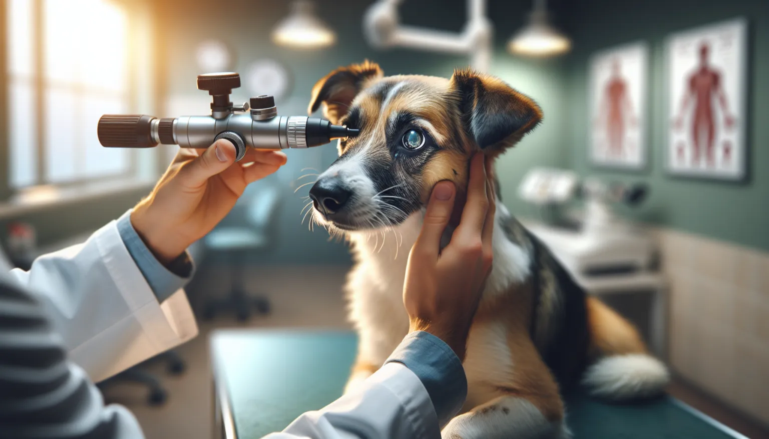
How Corneal Ulcers Are Diagnosed
Fluorescein stain is the key diagnostic tool — a dye that adheres to areas where the corneal epithelium is absent and fluoresces bright green under blue light. The staining pattern reveals ulcer size, depth, and location. A Schirmer tear test measures tear production to identify dry eye as an underlying cause. Intraocular pressure measurement checks for concurrent glaucoma.
Ulcer depth assessment is critical for treatment decisions. Superficial ulcers affecting only the epithelium are managed medically. Deep stromal ulcers (greater than 50% of stromal depth) and descemetoceles (where only Descemet’s membrane remains) require surgical intervention within hours to prevent perforation.
- Fluorescein stain to confirm ulcer presence, size, shape, and depth
- Schirmer tear test to evaluate tear production and identify dry eye as a contributing cause
- Intraocular pressure to rule out concurrent glaucoma
- Slit lamp examination or corneal topography in referral settings for detailed depth assessment
- Corneal cytology or culture in suspected infected (melting) ulcers to guide antibiotic selection
Treatment Depends on Ulcer Type
Superficial ulcers are treated with topical broad-spectrum antibiotic drops (tobramycin, triple antibiotic, or ciprofloxacin) applied every 6-8 hours to prevent secondary bacterial infection. An Elizabethan collar prevents self-trauma during healing. Most superficial ulcers heal within 5-7 days.
Indolent ulcers (SCCED) require debridement procedures — grid keratotomy, superficial keratectomy, or diamond burr debridement — to allow epithelial adhesion. These ulcers will not heal with drops alone.
Deep stromal ulcers with bacterial infection (melting ulcers, characterized by a gelatinous, rapidly expanding appearance) are emergencies. They require intensive topical antibiotics (every 1-2 hours initially), autologous serum drops (which contain anti-protease factors that limit enzymatic stromal destruction), and surgical consultation for conjunctival flap or corneoconjunctival transposition. Refer to a veterinary ophthalmologist for all cases with stromal depth beyond 50% or evidence of active corneal melting.
Key treatment rules:
- E-collar must be worn continuously until ulcer healing is confirmed by fluorescein re-stain
- Never use corticosteroid eye drops in a dog with a corneal ulcer — they delay healing and promote infection
- Recheck with fluorescein stain at 5-7 days to confirm healing — do not assume improvement based on behavioral signs alone
- Refer to veterinary ophthalmologist for deep ulcers, melting ulcers, or non-healing indolent ulcers
- Address underlying causes: treat dry eye with cyclosporine drops, correct entropion surgically if present
12-Week Recovery Plan
- Weeks 1-2 (baseline lock-in): Confirm diagnosis and ulcer depth. Start a shared household log tracking eye comfort, squinting, discharge, E-collar compliance, and drop schedule adherence.
- Weeks 3-4 (adherence audit): Verify that every caregiver follows the same drop schedule. Identify missed-dose friction and solve it. Confirm the E-collar stays on.
- Weeks 5-6 (response checkpoint): Compare current eye comfort against baseline. If the ulcer has not healed by the 7-day recheck, escalate to debridement or specialist referral immediately.
- Weeks 7-8 (risk tightening): Predefine escalation thresholds for recurrence signs. Confirm after-hours emergency route for acute eye pain.
- Weeks 9-10 (resilience build): Address underlying causes (dry eye treatment, entropion correction planning). Schedule breed-appropriate ophthalmology screening.
- Weeks 11-12 (handoff to maintenance): Document long-term follow-up cadence. For brachycephalic breeds, establish annual ophthalmology screening.
The Pattern That Leads to Vision Loss
The most common mistake is underestimating speed. Corneal ulcers can look minor at breakfast and become surgical emergencies by dinner, especially in brachycephalic breeds.
A second failure: stopping the E-collar early because “the dog seems better.” Behavioral improvement does not equal healed epithelium. Only a negative fluorescein re-stain confirms healing. Dogs that rub a healing ulcer can re-traumatize it or introduce infection.
In breeds prone to recurrence, the third failure is never addressing the underlying cause. Treating each ulcer in isolation without managing dry eye, entropion, or excessive corneal exposure guarantees the cycle continues.
Nutritional Considerations
There is no specific dietary intervention for corneal ulcers. General nutritional status supports immune function and wound healing. Omega-3 fatty acid supplementation has mild anti-inflammatory properties that may support ocular surface health, though direct evidence for corneal ulcer healing is limited.
For dogs with dry eye as the underlying cause of recurrent ulcers, omega-3 supplementation at evidence-based doses is a reasonable supportive measure alongside topical cyclosporine therapy.
- Omega-3 Fish Oil for Dogs: Evidence, Dosing Context, and Safety
- Feeding Guide for Adult Dogs: Maintenance Nutrition Without Drift
- Vitamin E for Dogs provides antioxidant support for ocular tissue health
For evidence context and execution details, review:
Follow-Up and Monitoring
Corneal ulcers require structured follow-up to confirm healing and prevent recurrence:
- Fluorescein recheck at 5-7 days to confirm epithelial closure — healing cannot be assumed from behavioral improvement alone
- If the ulcer is not healing at 7 days, escalate to debridement or specialist referral
- For dry eye: Schirmer tear test every 6 months to assess ongoing tear production and adjust cyclosporine concentration
- Brachycephalic breeds: annual ophthalmology screening to assess eyelid conformation, tear film quality, and corneal health
For brachycephalic breeds with recurrent corneal disease, an established relationship with a veterinary ophthalmologist allows systematic management of the anatomical factors that drive ongoing risk.
When to Seek Emergency Care
Seek same-day emergency care for:
- Any dog with acute eye squinting that is not improving within 2-4 hours
- Visible protrusion of uveal tissue (dark tissue) through the cornea — this indicates perforation
- Collapse or liquefaction of the corneal surface (melting appearance) — this indicates an enzymatic emergency
- Complete inability to open the eye combined with profuse discharge
- Any brachycephalic dog with acute eye signs — these breeds escalate rapidly
Related Condition Pathways
Corneal Ulcer often overlaps with adjacent pathways that affect diagnosis timing, treatment burden, and long-term resilience:
- Dry Eye (KCS): inadequate tear production is a major driver of recurrent corneal ulceration.
- Glaucoma: concurrent glaucoma can complicate corneal ulcer management and must be identified.
- Brachycephalic Syndrome: brachycephalic ocular anatomy dramatically increases corneal disease risk and severity.
These resources help you plan and prepare. Diagnostic confirmation and treatment changes are clinical decisions that require veterinary oversight.
Related Breed Longevity Guides
Brachycephalic breeds face the highest corneal ulcer risk due to their ocular anatomy:
- French Bulldog Lifespan & Longevity Guide
- English Bulldog Lifespan & Longevity Guide
- Boxer Lifespan & Longevity Guide
Brachycephalic breed owners should know corneal ulcer signs and have a plan for same-day evaluation. Waiting overnight when an eye is acutely painful can allow preventable progression to deep ulceration.
Additional Breeds at Elevated Risk
Frequently Asked Questions
How long does a corneal ulcer take to heal in a dog?
Simple superficial ulcers typically heal within 5-7 days with appropriate antibiotic treatment and E-collar use. Indolent ulcers (SCCED) take longer — often 2-4 weeks — and may require debridement procedures. Deep ulcers with bacterial infection can worsen within 24 hours without intensive treatment.
Can I use human eye drops on my dog’s corneal ulcer?
No. Never apply over-the-counter human eye drops to a dog’s eye without veterinary guidance. Many human eye preparations contain preservatives or active ingredients harmful to dogs. Corticosteroid-containing drops are particularly dangerous for corneal ulcers as they promote infection and delay healing.
Why does my dog keep getting corneal ulcers?
Recurrent ulcers in brachycephalic breeds are typically driven by anatomical factors — exophthalmos, lagophthalmos, and eyelid abnormalities — that increase corneal exposure and vulnerability to trauma. In older non-brachycephalic dogs, indolent ulcers (SCCED) are a common cause. Dry eye (KCS) is another major recurrence driver. Identifying and treating the underlying cause is essential for breaking the recurrence cycle.
What is a melting ulcer?
A melting ulcer (keratomalacia) involves enzymatic destruction of the corneal stroma by bacterial proteases, producing a rapidly expanding gelatinous defect that can perforate within hours. It is a true ocular emergency requiring immediate intensive topical treatment and surgical consultation.
Is corneal ulcer surgery necessary?
Surgery is necessary for deep stromal ulcers, descemetoceles (Descemet’s membrane exposure), and corneal perforations. Surgical options include conjunctival flap placement, corneoconjunctival transposition, or corneoscleral transposition, which provide structural support and blood supply to the healing cornea. Referral to a veterinary ophthalmologist is recommended for surgical cases.
Medical Disclaimer
Corneal ulcers are painful ocular emergencies that can cause permanent vision loss if untreated or inadequately managed. Do not wait to seek veterinary evaluation for a dog with eye squinting or obvious ocular discomfort.
References
- Maggs DJ et al. Slatter’s Fundamentals of Veterinary Ophthalmology, 5th ed. Saunders. 2013.
- Williams DL. Canine chronic corneal epithelial defects. Vet Clin North Am Small Anim Pract. 2008.
- Ledbetter EC, Gilger BC. Diseases and surgery of the canine cornea and sclera. In: Veterinary Ophthalmology, 5th ed. 2013.
- Crispin SM. Corneal diseases of the dog. Vet Rec. 1989.
- ACVO Genetics Committee. Ocular disorders presumed to be inherited in purebred dogs. 2015.
Related reads
Related Reading
Continue exploring