Evidence deep dives for Cutaneous Histiocytoma
Pair mechanism-level evidence with practical protocol context before discussing next steps with your veterinarian.
The Bump That Appeared Overnight
A small, pink bump appears on your young dog’s ear seemingly overnight. Within two weeks it doubles in size. The natural reaction is alarm — but in most cases, this is a cutaneous histiocytoma, one of the most common and most benign skin tumors in dogs.
Histiocytomas arise from epidermal Langerhans cells, a type of immune cell normally found in the skin. They grow fast, look concerning, and then — in the majority of cases — disappear on their own within one to three months as the immune system mounts a targeted T-cell response and destroys them.
A typical histiocytoma appears solitary, dome-shaped or button-like, smooth-surfaced and hairless, measuring 1-3 cm across. It tends to be pink to red (reflecting its highly vascular, cellular nature) and may ulcerate on the surface, particularly as regression begins.
That rapid appearance — a new mass that seems to come from nowhere over days to weeks — is characteristic and does not indicate malignancy in this tumor type.
Here is the critical distinction owners need to understand: while histiocytomas are almost always benign, they can look identical to mast cell tumors on the surface. Mast cell tumors carry a dramatically different prognosis. No skin mass should be assumed benign based on appearance alone.
The Longevity Impact
The longevity relevance of a histiocytoma is diagnostic, not prognostic. The tumor itself poses no threat. The danger lies in misidentification.
An owner or clinician who assumes a mass is “probably a histiocytoma” and waits for regression may be watching a mast cell tumor, sarcoma, or other aggressive cancer grow unchecked. The observation window that is safe for a confirmed histiocytoma can allow a malignant tumor to become locally invasive or metastatic.
For confirmed histiocytomas, the outlook is excellent. Spontaneous resolution typically occurs within one to three months, and the dog’s long-term health remains unaffected. Multiple concurrent histiocytomas or new masses in older dogs warrant closer investigation.
Signs and Recognition
Histiocytomas have a characteristic appearance, but “characteristic” does not mean “pathognomonic” — other tumors can look the same.
What to watch for:
- A rapidly appearing, dome-shaped or button-like skin mass, usually 1-3 cm at presentation
- Smooth, pink to red surface, often hairless or with thinned hair over the mass
- Found most often on the head (especially ears and face), neck, and limbs
- Usually a single mass; occasionally two or three simultaneous lesions
- Surface ulceration, particularly as regression begins
- Generally not painful unless ulcerated or traumatized
- Rapid growth from first appearance — often doubled in size within 2-3 weeks
Even if this description fits perfectly, get cytology before deciding to watch and wait. Mast cell tumors can look identical on gross examination.
The Diagnostic Process
Fine-needle aspiration cytology is the primary diagnostic test, and it is usually sufficient. A veterinarian inserts a small needle into the mass, collects cells, and examines them under a microscope. Histiocytoma cytology reveals sheets of round cells with pale cytoplasm and reniform (kidney-shaped) nuclei. The absence of mast cell granules — which appear as purple-staining dots on Diff-Quik stain — supports histiocytoma over mast cell tumor. In experienced hands, cytology is highly accurate for this tumor type.
Histopathology of an excised sample provides definitive diagnosis. It is warranted when cytology is inconclusive, the mass does not behave as expected (no regression within 8-12 weeks), or when malignant differentials cannot be ruled out. Immunohistochemistry for Langerhans cell markers (CD1a, CD11c, MHC class II) can confirm histiocytic origin in ambiguous cases.
The diagnostic sequence:
- Fine-needle aspiration cytology: the most practical first step — perform on any new skin mass before choosing observation
- Diff-Quik staining: specifically examine for mast cell granules to exclude mast cell tumor
- Histopathology if cytology is inconclusive or the mass fails to regress within 8-12 weeks
- Immunohistochemistry for CD1a and Langerhans cell markers if histopathology remains ambiguous
Treatment and Management
Most confirmed histiocytomas need no treatment at all. The immune system handles it. The owner’s job during the observation period is structured monitoring, not intervention.
Counsel your household to monitor the mass at home with weekly photographs and report any change in size trajectory, ulceration, or signs of discomfort.
Surgical excision makes sense in specific situations: when spontaneous regression has not occurred within three months, when the mass sits in a location that causes self-trauma (face, feet), when ulceration leads to secondary infection, or when the owner simply prefers definitive removal. Excision is generally curative — recurrence after complete removal is uncommon.
A few important notes on what not to do. Corticosteroids are not routinely indicated and may actually delay regression by suppressing the T-cell response driving resolution. Antibiotics address secondary bacterial infection in ulcerated masses, but gentle topical care (keeping the area clean and dry) often suffices for mild ulceration.
Practical management steps:
- Confirm diagnosis with fine-needle aspiration cytology before choosing watchful waiting
- Monitor with weekly photographs and a measurement log
- Prevent self-trauma with an Elizabethan collar if the dog licks or rubs the mass
- Schedule surgical excision if the mass has not significantly regressed within 8-12 weeks
- Treat ulcerated surfaces with gentle antiseptic cleaning and topical antibiotics if secondary infection develops
12-Week Monitoring and Response Plan
- Weeks 1-2 (baseline lock-in): Confirm the cytological diagnosis. Start a shared household log with weekly photographs, measurements, and notes on the dog’s comfort, appetite, and activity level.
- Weeks 3-4 (adherence audit): Verify that everyone in the household follows the same monitoring protocol. Identify any friction in the photo/measurement routine and fix it.
- Weeks 5-6 (response checkpoint): Compare current mass size and surface appearance against baseline photos. If no regression trend is visible, flag this for your veterinarian rather than continuing to wait passively.
- Weeks 7-8 (risk tightening): Predefine escalation thresholds. Know exactly what changes — rapid growth resumption, new masses, systemic signs — should trigger immediate veterinary contact rather than continued observation.
- Weeks 9-10 (resilience build): If regression is underway, maintain routine monitoring. If not, pursue surgical consultation as planned.
- Weeks 11-12 (handoff to maintenance): Document outcome. If the mass resolved, no further specific monitoring is needed beyond routine wellness visits. If multiple histiocytomas occurred or the dog is older, discuss systemic screening.
Most-Missed Drift Pattern
The biggest mistake with histiocytomas is not the tumor itself — it is the process around it.
Families commonly react only to dramatic changes and miss the subtler signals that something is off. A mass that plateaus instead of shrinking at week six deserves attention, not another month of passive observation. Conversely, owners sometimes over-intervene: rushing to surgery on a mass that cytology confirms as benign and that is already beginning to regress.
The most common process failure across multi-caregiver households is inconsistent execution. When one person photographs weekly and another does not, trend data becomes unreliable and veterinary decisions lose their foundation.
Teams that review one objective metric each week — mass diameter, surface condition, dog comfort — detect problems far earlier than those who rely on general impressions.
Nutritional Considerations
No specific nutritional interventions are needed for cutaneous histiocytoma. General nutritional adequacy supports the immune function responsible for spontaneous regression. Maintaining lean body condition and feeding a complete, balanced diet gives the immune system what it needs.
For dogs undergoing surgical excision, adequate protein intake supports wound healing. No supplements are indicated specifically for this condition.
- Feeding Guide for Adult Dogs: Maintenance Nutrition
- Multi-Vitamin for Dogs: Evidence Review
- Omega-3 Fish Oil for Dogs supports general immune function relevant to the T-cell response that drives spontaneous regression
For evidence context and execution details, review:
- Canine Cancer Early Warning Workflow
- Genetic Testing for Dogs: Clinical ROI
- Cancer Screening in Dogs: What Helps
Monitoring and Follow-Up
Structured observation of a confirmed histiocytoma follows a clear timeline:
- Weekly photographs from consistent angles and distance to document size trajectory objectively
- Weekly measurement using a ruler placed adjacent to the mass
- Report any sudden rapid growth resumption, change in consistency, or failure to shrink by 4-6 weeks
- Veterinary recheck at 6-8 weeks if spontaneous regression has not begun
- Surgical consultation at 8-12 weeks for non-regressing masses
Once a confirmed histiocytoma completes spontaneous regression, no further specific monitoring is needed beyond routine wellness visits. Dogs with multiple concurrent histiocytomas or recurrence at the same site warrant more thorough investigation, including screening for systemic histiocytic disease.
When to Contact Your Veterinarian
Reach out promptly for:
- Rapid resumed growth after an initial plateau or shrinkage — this may indicate incorrect initial diagnosis or secondary change
- Mass failure to regress by 10-12 weeks after cytological confirmation
- New masses developing simultaneously in other locations or in regional lymph nodes
- Systemic signs (lethargy, weight loss, reduced appetite) in a dog with multiple skin masses
- Severe or worsening ulceration with active infection that does not respond to topical care
Related Condition Pathways
Cutaneous Histiocytoma often overlaps with adjacent pathways that affect diagnosis timing, treatment burden, and long-term resilience:
- Mast Cell Tumor: mast cell tumors are the most critical differential for cutaneous histiocytoma — cytology differentiates them; never observe without cytology first.
- Skin Cancer (General): histiocytoma is a benign skin tumor, but any new skin mass warrants cytological assessment before assuming benign nature.
- Cancer (General): rarely, systemic histiocytic sarcoma — a malignant histiocytic cancer — can produce multiple skin lesions; multiple simultaneous histiocytomas warrant systemic evaluation.
These guides provide background for productive veterinary conversations — they do not replace clinical evaluation or treatment planning.
Related Breed Longevity Guides
Cutaneous histiocytoma shows breed predisposition, with retriever-type and brachycephalic breeds overrepresented:
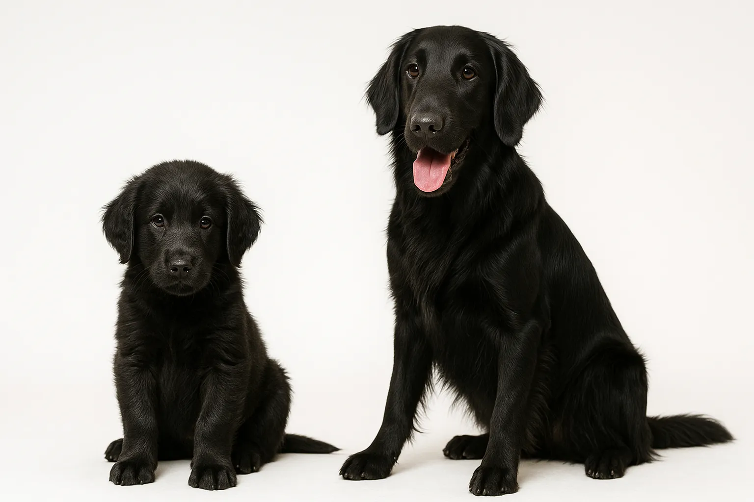
Flat-Coated Retrievers, who carry an overall elevated tumor burden, have disproportionately high histiocytoma incidence. Boxers are notable for high rates of multiple skin tumor types, with histiocytoma among the most common benign forms in this breed.
Frequently Asked Questions
Should I have a histiocytoma removed or wait?
If cytology confirms a histiocytoma, watchful waiting for 1-3 months is reasonable in most cases, as the majority regress spontaneously. Surgical removal is recommended if the mass has not regressed within 8-12 weeks, if it is in a location causing self-trauma, or if secondary infection from ulceration is recurring. Either approach is medically sound for a confirmed histiocytoma.
How do I know it’s not a mast cell tumor?
Fine-needle aspiration cytology with Diff-Quik staining can distinguish the two in most cases. Mast cell tumors contain characteristic purple-staining granules visible on cytology; histiocytomas do not. Any new skin mass, regardless of appearance, should have cytology performed before a “wait and watch” approach is adopted. Mast cell tumors can look identical to histiocytomas on gross examination.
Can histiocytomas come back after removal?
Recurrence of a confirmed histiocytoma after complete surgical excision is uncommon. If a new mass appears at the same site after excision, or if multiple simultaneous histiocytomas develop, investigation for systemic histiocytic disease (which involves malignant histiocytes and has a different prognosis) should be undertaken.
Are histiocytomas more common in certain breeds?
Yes. Labrador Retrievers, Golden Retrievers, Flat-Coated Retrievers, Boxers, and English Cocker Spaniels are overrepresented in case series. Young dogs (under 3 years) are most commonly affected. The condition is rare in dogs over 6 years, and new masses in older dogs should raise higher suspicion for malignant differentials.
What if my dog has multiple histiocytomas at once?
Multiple simultaneous cutaneous histiocytomas can occur but are less typical than solitary lesions. They warrant cytological or histopathological confirmation from multiple sites and, if all confirm benign histiocytoma histology, careful monitoring for spontaneous regression. The development of many simultaneous histiocytic-appearing lesions in an adult or older dog raises the concern for reactive histiocytosis or early systemic histiocytic disease, which require veterinary oncology input.
Medical Disclaimer
This content is for educational purposes only and does not constitute veterinary medical advice. Any new skin mass should be evaluated by a veterinarian and assessed with fine-needle aspiration cytology before a watchful-waiting approach is adopted. Do not assume a mass is benign based on appearance alone, as mast cell tumors and other malignant tumors can closely mimic cutaneous histiocytoma.
References
- Mays MB, Bergeron JA. Cutaneous histiocytomas of the dog. J Am Vet Med Assoc. 1986;188(4):377-381.
- Fulmer AK, Mauldin GE. Canine histiocytic neoplasia: an overview. Can Vet J. 2007;48(10):1041-1043.
- Moore PF. A review of histiocytic diseases of dogs and cats. Vet Pathol. 2014;51(1):167-184.
- Wiley JL, Zerbe CA, Brodey RS. Histiocytoma in dogs: a retrospective study of 37 cases (1979-1988). J Am Anim Hosp Assoc. 1990;26:617-624.
- Affolter VK, Moore PF. Localized and disseminated histiocytic sarcoma of dendritic cell origin in dogs. Vet Pathol. 2002;39(1):74-83.
Related reads
Related Reading
Continue exploring