Evidence deep dives for Discoid Lupus Erythematosus
Pair mechanism-level evidence with practical protocol context before discussing next steps with your veterinarian.
When the Nose Starts Losing Its Color
A dog’s nose changes color. The dark, textured surface fades to gray, then pink. Small cracks and sores appear. The owner assumes it is sunburn or dry skin. Months pass before anyone realizes the immune system is attacking its own tissue.
Discoid lupus erythematosus (DLE) is the most common immune-mediated skin disease in dogs. It targets the skin — primarily the nose, face, and occasionally the lips, periorbital area, and ears — without the multi-organ damage that characterizes systemic lupus erythematosus (SLE). This distinction matters enormously: DLE carries a far better prognosis and rarely progresses to systemic disease.
The hallmark of DLE is immune-mediated destruction at the dermal-epidermal junction, concentrated on the nasal planum. A healthy dog’s nose has a characteristic cobblestone texture and dark pigmentation. In DLE, the immune attack strips away that pigment, erases the cobblestone pattern, and opens surface erosions that can deepen into painful ulcerations. In advanced cases, the nose becomes smooth, pale, and prone to cracking.
Ultraviolet light is a major accelerant. Dogs exposed to direct sunlight experience faster lesion progression than those kept out of UV. This photosensitivity is both a defining clinical feature and a management lever — controlling UV exposure is one of the most effective things an owner can do.
DLE is not contagious. No pathogen causes it, and it cannot spread between dogs. Certain breeds carry a strong heritable predisposition, though researchers have not yet fully mapped the specific genetics.
Why This Matters for Your Dog’s Healthspan
DLE reduces quality of life primarily through chronic nasal pain, disfigurement, and the risk of ulceration progressing to secondary infection. Without treatment, DLE can produce severe nasal erosions that expose underlying cartilage, cause significant discomfort, and dramatically raise susceptibility to bacterial infection at an anatomically vulnerable site.
The key longevity consideration is that chronic, uncontrolled UV-induced tissue damage at DLE lesion sites carries a low but real risk of malignant transformation over time. Chronically sun-damaged, depigmented tissue faces higher susceptibility to squamous cell carcinoma — a serious complication that appropriate ongoing management can largely prevent.
Dogs with confirmed DLE should also be periodically evaluated to exclude progression toward systemic lupus, which involves kidney, joint, and blood system changes. This transition is uncommon, but annual screening in actively affected animals is prudent.
Early Signs and Recognition
DLE lesions almost always start on the nose. Early recognition can prevent months of unnecessary progression:
- Loss of the normal cobblestone texture, replaced by a smooth surface
- Early depigmentation: gray, pink, or white patches developing on a previously dark nose
- Mild erosions or superficial crusting on the nasal planum
- The nose appearing redder or more inflamed after sun exposure
- Occasional nosebleeds from surface erosion
- Depigmentation extending to the nasal bridge or lip margins in more advanced cases
A characteristic seasonal pattern — worsening in summer, improvement in winter — combined with breed predisposition should raise strong clinical suspicion even before biopsy confirms the diagnosis.
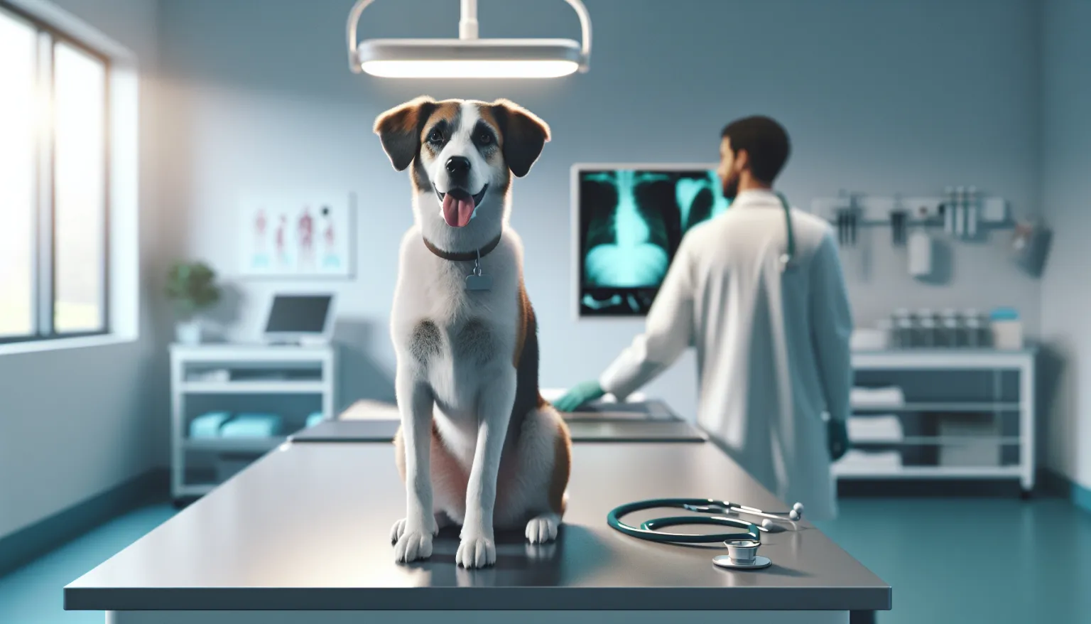
Getting to a Diagnosis
Definitive diagnosis requires a skin biopsy of the nasal planum. Histopathology reveals hydropic degeneration of basal keratinocytes, subepidermal vacuolation, and a lichenoid interface dermatitis with lymphocytic infiltration. This pattern distinguishes DLE from systemic lupus, deep fungal infections (which can mimic DLE), and nasal pyoderma. Biopsy should be performed under sedation or general anesthesia to get adequate tissue depth.
Direct immunofluorescence of biopsy samples demonstrates immunoglobulin and complement deposition at the dermal-epidermal junction in approximately 60-70% of DLE cases, supporting the immune-mediated mechanism.
Antinuclear antibody (ANA) testing is important to exclude systemic lupus. A positive ANA with multi-organ signs changes both diagnosis and management significantly.
The diagnostic sequence:
- Punch biopsy of the nasal planum (3-4 mm, from the margin of active lesions)
- Histopathology identifying interface dermatitis with basal cell vacuolation
- Direct immunofluorescence for immunoglobulin deposition at the basement membrane zone
- ANA titer to screen for systemic lupus
- CBC and urinalysis to screen for systemic involvement (hemolytic anemia, proteinuria)
- Culture of nasal erosions if secondary bacterial infection is suspected
Treatment and Long-Term Management
Sun avoidance is the most impactful non-pharmacological intervention. Dogs with DLE should avoid peak UV hours (10 AM to 4 PM), stay out of direct sunlight during outdoor time, and have high-SPF sunscreen (SPF 30 or higher, formulated for dogs or free of zinc oxide and PABA) applied to the nasal planum before any UV exposure. This single measure can substantially reduce flare frequency and lesion severity.
Topical tacrolimus 0.1% ointment, applied twice daily, is the first-line pharmacological treatment for mild to moderate DLE. It suppresses T-cell activity locally without systemic immunosuppression. Response typically appears within 4-8 weeks. Topical corticosteroids (triamcinolone, betamethasone) work for acute flares but should not be used long-term on the nasal planum due to skin atrophy risk.
For dogs with significant erosions, pain, or poor topical response, systemic options include prednisone (initial 1-2 mg/kg daily, tapered over 8-12 weeks) or the combination of doxycycline and niacinamide. The doxycycline/niacinamide approach has shown efficacy in some dogs without the side effect burden of corticosteroids and is often tried before committing to systemic steroids in moderate cases.
Daily management checklist:
- Apply dog-safe sunscreen (no zinc oxide) to the nasal planum before any outdoor UV exposure
- Limit outdoor activity during peak UV hours year-round, not just in summer
- Apply topical tacrolimus 0.1% twice daily; wear gloves to prevent owner absorption
- Monitor nasal planum erosions monthly with photographs from consistent angles
- Treat secondary bacterial nasal infections promptly with appropriate antibiotics
Getting Started: The First 12 Weeks
- Weeks 1-2 (baseline lock-in): Confirm the biopsy diagnosis. Start a shared household log capturing daily markers: nasal condition, appetite, activity tolerance, any medication side effects.
- Weeks 3-4 (adherence audit): Verify that every caregiver follows the same sunscreen, medication, and UV-avoidance protocol. Identify the one step most likely to be skipped and build a fix.
- Weeks 5-6 (response checkpoint): Compare current nasal photographs against baseline. If erosions have not stabilized or improved, escalate to your veterinarian rather than waiting longer.
- Weeks 7-8 (risk tightening): Predefine what worsening looks like — deepening ulcers, new lesion locations, systemic signs — so that urgent decisions are never improvised.
- Weeks 9-10 (resilience build): Reinforce UV-avoidance habits and medication consistency as permanent routines, not temporary fixes.
- Weeks 11-12 (handoff to maintenance): Document the long-term reassessment cadence. Schedule the next veterinary check. Decide which metrics stay in weekly tracking.
Most-Missed Drift Pattern
The most common failure in DLE management is responding only to severe flares while ignoring the slow drift that precedes them. A nose that was stable in January and slightly worse in March is drifting — and the earlier that drift triggers reassessment, the less aggressive the correction needs to be.
Inconsistent household execution is the second most common problem. When one person applies sunscreen and another forgets, when medication timing varies by hours each day, trend data becomes noise. The disease appears “unpredictable” when in reality the management is inconsistent.
Teams that review one objective metric weekly — even just a consistent-angle photo of the nasal planum — detect relapse far earlier than those relying on general impressions.
Nutritional and Supplement Considerations
Omega-3 fatty acid supplementation (EPA and DHA from fish oil) provides systemic anti-inflammatory support that may help modulate immune hyperactivity relevant to DLE. No DLE-specific studies exist in dogs, but the immune-modulating effects of omega-3s are well characterized across dermatological immune conditions.
Vitamin E has antioxidant properties that theoretically protect UV-damaged skin. Some dermatologists include oral vitamin E (400-600 IU daily) as adjunct therapy for photosensitive conditions, though direct evidence in canine DLE remains limited.
Niacinamide (vitamin B3), used systemically in combination with doxycycline, also has topical skin-protecting properties.
- Omega-3 Fish Oil for Dogs: Evidence and Dosing
- Vitamin E for Dogs: Safety and Evidence
- Feeding Guide for Adult Dogs for baseline nutritional support during long-term immunomodulatory therapy
For evidence context and execution details, review:
- Genetic Testing for Dogs: Clinical ROI
- Supplement Evidence for Dog Longevity
- Spay-Neuter Timing and Longevity Context
Monitoring and Response Assessment
Long-term DLE monitoring centers on lesion control and preventing progression:
- Monthly nasal planum photography from a fixed angle and distance to track pigmentation and erosion changes objectively
- Semi-annual veterinary skin examination with ANA titer if systemic signs develop
- Evaluation for early squamous cell carcinoma at each exam when chronic depigmentation or erosion is present
- Seasonal tracking of UV exposure patterns with corresponding adjustment of protection measures
DLE is a manageable chronic condition in most dogs. With consistent sun protection and appropriate topical therapy, the prognosis for quality of life is good. A minority of cases require periodic escalation to systemic therapy.
When to Contact Your Veterinarian
Reach out promptly for:
- Nasal erosions deepening into ulcers with exposed cartilage or active bleeding
- Recurrent or heavy nosebleeds
- New lesions developing beyond the nose — lips, periorbital skin, ears, genitalia
- Signs of systemic involvement: joint swelling, lethargy, reduced appetite, or blood in urine
- A rapidly changing nasal planum lesion, particularly one that becomes raised, firm, or fails to heal (warrants biopsy to exclude malignant transformation)
Related Condition Pathways
Discoid Lupus Erythematosus often overlaps with adjacent pathways that affect diagnosis timing, treatment burden, and long-term resilience:
- Skin Cancer: chronic depigmented, UV-exposed nasal tissue has elevated squamous cell carcinoma risk — periodic biopsy warranted if lesions change character.
- Skin Allergies (Atopic Dermatitis): allergic skin disease may occur concurrently; differentiated by histopathology and lesion distribution.
- Hypothyroidism: thyroid dysfunction can worsen skin immune regulation and should be excluded in affected dogs.
These guides provide background for productive veterinary conversations — they do not replace clinical evaluation or treatment planning.
Related Breed Longevity Guides
Discoid lupus erythematosus shows clear breed predisposition, particularly in herding and working breeds:
German Shepherds are most frequently represented in DLE case series. Dogs with lighter nose pigmentation or those living at high altitude (greater UV index) face greater lesion burden regardless of breed.
Frequently Asked Questions
Will discoid lupus erythematosus progress to systemic lupus?
Progression from DLE to systemic lupus erythematosus is uncommon. The two conditions share immune dysregulation mechanisms but DLE is generally considered a distinct, skin-limited disease. Dogs with DLE should have annual ANA testing and urinalysis to monitor for early systemic involvement, but most affected dogs never develop SLE.
Can the nose pigmentation return after DLE treatment?
Partial repigmentation can occur, particularly in dogs treated early before significant gland and pigment cell destruction. Complete return to original nasal color is uncommon in dogs with established lesions. The primary treatment goal is halting erosion progression and maintaining skin integrity, not necessarily restoring full pigmentation.
How important is sun avoidance in DLE management?
Extremely important. Ultraviolet light is a primary trigger for DLE exacerbation, and dogs with uncontrolled UV exposure show significantly faster lesion progression. Sun avoidance and UV-protective sunscreen are foundational — pharmaceutical treatment alone without UV management provides incomplete disease control.
Is topical tacrolimus safe for long-term use on the nose?
Topical tacrolimus has a good safety profile for long-term dermatological use in dogs. The main concern is owner absorption through skin contact — handlers should wear gloves when applying. Systemic absorption from nasal application in dogs is low. Annual monitoring is prudent in dogs on long-term therapy.
Can DLE lesions become cancerous?
Chronically sun-damaged, depigmented nasal tissue has elevated risk for squamous cell carcinoma development compared to normal skin. This risk is low but real. Any DLE lesion that changes character — becomes raised, firm, proliferative, or fails to heal — warrants biopsy to exclude malignant transformation.
What sunscreen can I use on my dog’s nose?
Use pet-specific sunscreens or human sunscreens that are free of zinc oxide and para-aminobenzoic acid (PABA), both of which are toxic to dogs if licked. SPF 30 or higher is recommended. Apply before outdoor exposure and reapply after swimming, rain, or if the dog licks the nose. Ask your veterinarian for specific product recommendations.
Medical Disclaimer
This content is for educational purposes only and does not constitute veterinary medical advice. Discoid lupus erythematosus requires professional diagnosis via skin biopsy to distinguish it from other nasal diseases including infection, immune-mediated disease, and nasal neoplasia. Treatment should be supervised by a veterinary dermatologist or veterinarian experienced with immune-mediated skin conditions.
References
- Bryden SL, White SD, Dunston SM, et al. Clinical, histopathological, and immunological characteristics of exfoliative cutaneous lupus erythematosus in 25 German Short-Haired Pointers. Vet Dermatol. 2005;16(4):239-252.
- Scott DW, Miller WH Jr. Mucocutaneous lupus erythematosus in the dog: a retrospective analysis of 10 cases (1983-2005). Jpn J Vet Dermatol. 2007;13(3):139-150.
- Olivry T, Linder KE. Dermatoses affecting desmosomes in animals: a mechanistic review of acantholytic blistering skin diseases. Vet Dermatol. 2009;20(5-6):313-326.
- Mauldin EA, Peters-Kennedy J. Integumentary system. In: Maxie MG, ed. Jubb, Kennedy and Palmer’s Pathology of Domestic Animals. 6th ed. Elsevier; 2016:509-736.
- Griffies JD, Mendelsohn CL, Rosenkrantz WS, et al. Topical 0.1% tacrolimus for the treatment of discoid lupus erythematosus and pemphigus erythematosus in dogs. J Am Anim Hosp Assoc. 2004;40(1):29-41.
Related reads
Related Reading
Continue exploring