Evidence deep dives for Ectropion
Pair mechanism-level evidence with practical protocol context before discussing next steps with your veterinarian.
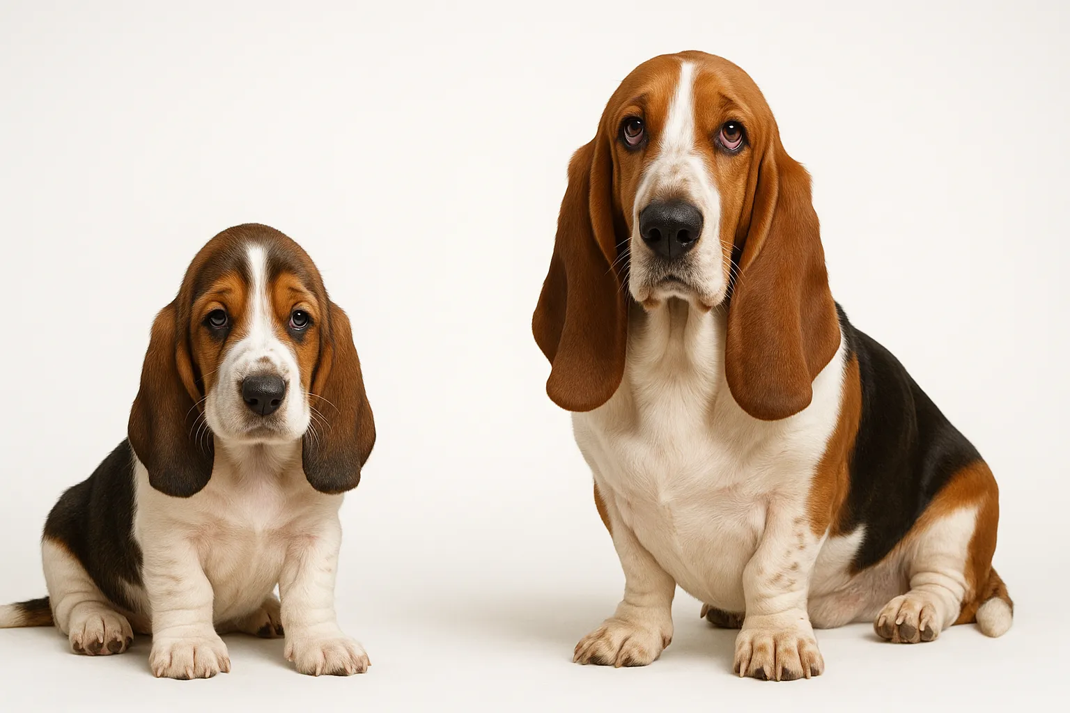
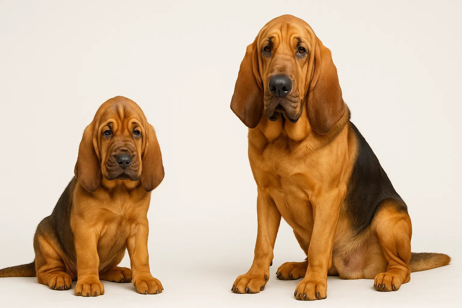
Those Soulful Droopy Eyes Come With a Built-In Vulnerability
If you own a Bloodhound, Basset Hound, or Saint Bernard, you have likely admired those droopy, soulful eyes. What you may not realize is that those eyes come with a built-in vulnerability — ectropion.
Ectropion is the outward rolling or drooping of the lower eyelid, exposing the conjunctival lining (the pink tissue inside the eyelid) and creating a pocket between the eyelid and the eyeball. This pocket collects debris, allergens, mucus, and bacteria that would normally be swept away by the blinking eyelid. The exposed conjunctiva dries out and becomes chronically inflamed.
Unlike entropion, where the eyelid rolls inward and abrades the cornea, ectropion creates the opposite structural problem — insufficient eyelid-to-globe contact. Both conditions are common in breeds selected for loose facial skin, and some dogs have “diamond eye” where the upper lid shows entropion while the lower lid shows ectropion simultaneously.
Three forms exist: conformational (breed-related, present from birth), acquired (from scarring, nerve damage, or aging), and cicatricial (resulting from previous surgery, including overcorrection of entropion). The conformational form accounts for the vast majority of cases.
Beyond Treatment: The Longevity Dimension
Ectropion alone rarely threatens life. But its chronic nature creates a persistent cycle of conjunctivitis, corneal exposure, and secondary infection that chips away at quality of life over years. Dogs with untreated moderate to severe ectropion endure daily ocular discomfort — eye discharge matting the facial fur, recurrent bacterial conjunctivitis demanding antibiotic courses, and progressive corneal damage from exposure keratitis.
The longevity concern is not mortality — it is morbidity. A dog living with constant discomfort, frequent eye medications, and recurrent infections has a measurably reduced quality of life. Managing ectropion proactively prevents this slow accumulation of ocular damage.
Early Signs and Home Monitoring
Ectropion is usually visible on inspection, but the secondary complications are what bring most owners to the veterinarian:
- visible drooping of the lower eyelid, exposing pink conjunctival tissue
- chronic mucoid or mucopurulent discharge accumulating in the lower eyelid pocket
- tear staining on the face below the affected eye
- redness and swelling of the exposed conjunctiva
- frequent squinting, blinking, or pawing at the eyes
- crusting of discharge along the eyelid margins
- recurrent bacterial conjunctivitis requiring repeated antibiotic treatment
- corneal cloudiness or vascularization in advanced cases (from chronic exposure)
In breeds with naturally loose facial skin, the distinction between “normal breed conformation” and clinically significant ectropion requires veterinary assessment. Not every droopy eyelid needs surgery, but every droopy eyelid needs monitoring.
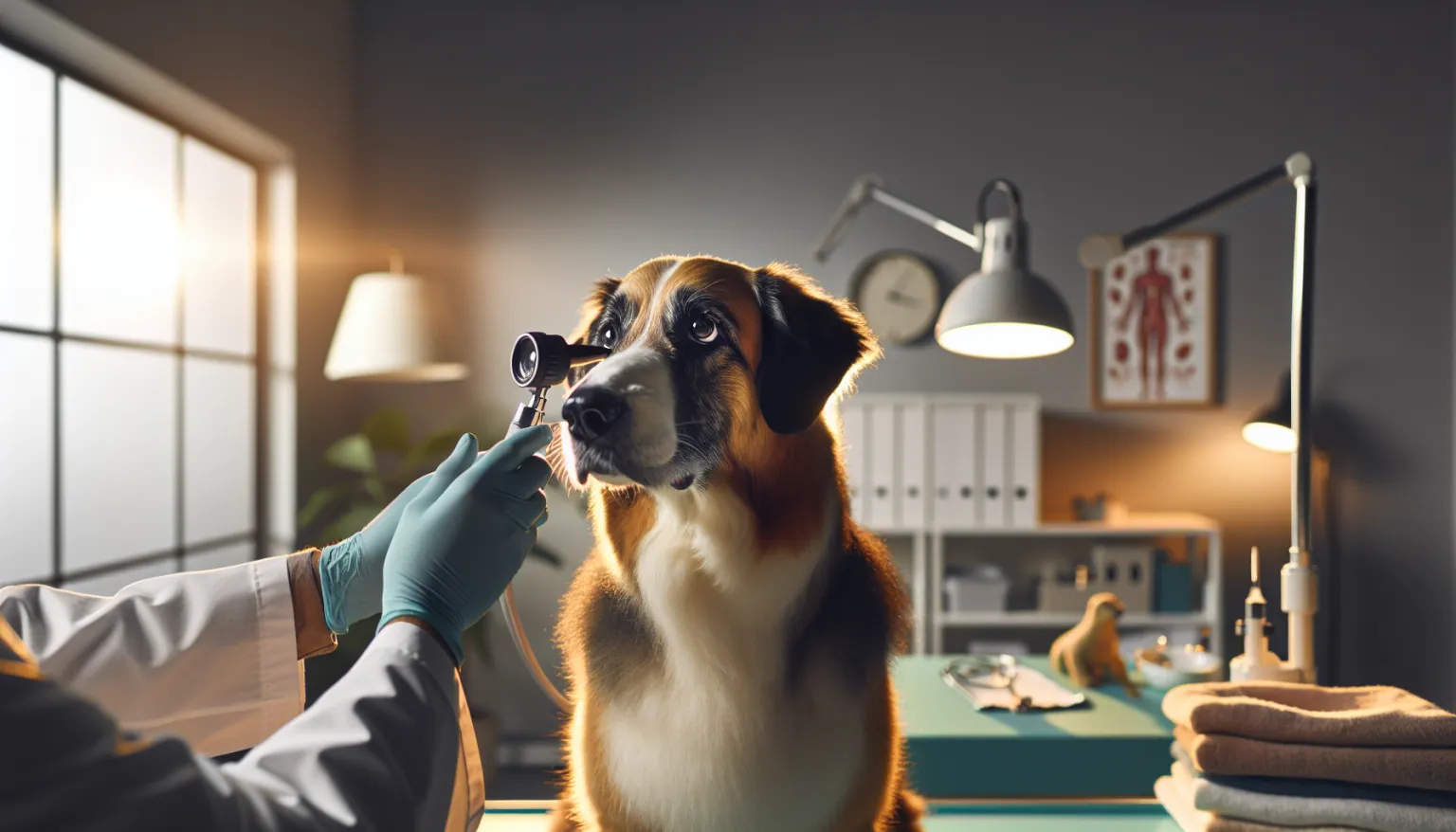
How Ectropion Is Diagnosed
Diagnosis is based on clinical examination of eyelid position and assessment of secondary complications. Your veterinarian evaluates:
- the degree of lower eyelid eversion and the depth of the conjunctival pocket
- whether the condition is unilateral or bilateral
- the presence and severity of conjunctivitis
- corneal integrity using fluorescein staining to detect ulceration
- tear production using the Schirmer Tear Test — chronic exposure can alter tear dynamics
- whether entropion coexists with ectropion (the “diamond eye” configuration)
Key diagnostic considerations:
- distinguish conformational ectropion from acquired or cicatricial forms — the underlying cause determines whether surgery is appropriate
- assess corneal health thoroughly before any anesthetic procedure
- in older dogs with new-onset ectropion, evaluate for cranial nerve deficits or neuromuscular disease
- document baseline eyelid position photographically for longitudinal comparison
Breeds at Risk
Ectropion is a conformational condition strongly linked to breeds selected for loose facial skin and heavy facial folds:
- Bloodhound — among the most commonly and severely affected breeds
- Basset Hound — prominent ectropion is virtually universal in the breed
- English Mastiff — heavy facial skin contributes to lower lid laxity
- Saint Bernard — large skull and loose facial skin predispose strongly
- Great Dane — especially harlequin and mantle varieties
- Neapolitan Mastiff — extreme skin laxity affects multiple periocular structures
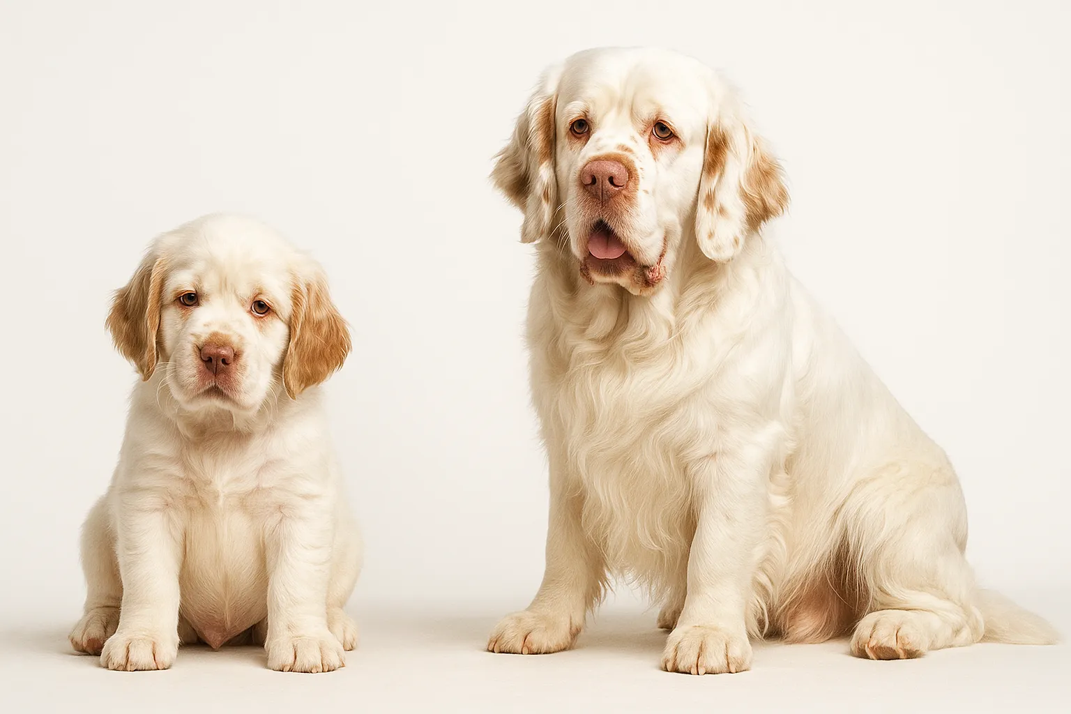
- Clumber Spaniel — breed standard acknowledges the droopy lower lid
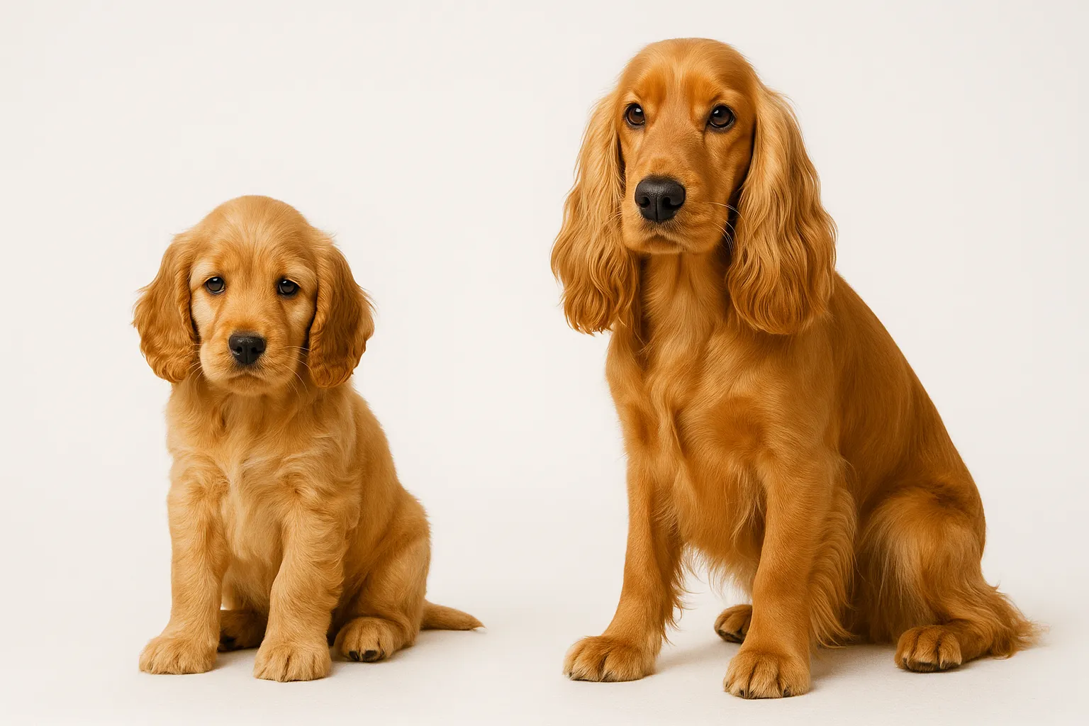
- Cocker Spaniel — lower lid laxity is common, often mild
Giant breeds and breeds with heavy facial conformation are disproportionately affected. In many of these breeds, some degree of ectropion is considered a normal breed characteristic, which makes the line between “normal for the breed” and “clinically problematic” a judgment call that requires ophthalmologic expertise.
Prevention Strategies
Conformational ectropion cannot be prevented in an individual dog — it is an inherited structural trait embedded in breed conformation. The only population-level prevention is selective breeding away from extreme facial skin laxity, which conflicts with current breed standards in several affected breeds.
What owners can control is preventing the complications of ectropion:
- daily eye cleaning to remove accumulated debris from the conjunctival pocket
- lubricating eye drops (artificial tears) to protect exposed conjunctival tissue and cornea
- prompt treatment of bacterial conjunctivitis before it becomes chronic
- protecting eyes from wind, dust, and environmental irritants during outdoor activities
- regular veterinary eye examinations to catch corneal changes early
- avoiding exposure to known ocular irritants (dusty environments, chemical sprays)
Treatment and Long-Term Management
Treatment depends on the severity of ectropion and the presence of secondary complications.
Mild ectropion (cosmetic, minimal symptoms):
Many dogs with mild breed-related ectropion require no treatment beyond routine eye care. Regular cleaning and monitoring is sufficient.
Moderate ectropion (chronic conjunctivitis, recurrent infections):
- lubricating eye drops 2-3 times daily to protect the exposed tissue
- topical antibiotics for acute bacterial conjunctivitis episodes
- daily eye cleaning with sterile saline or veterinary eye wash
- environmental modification to reduce irritant exposure
- referral for surgical evaluation if medical management is insufficient
Severe ectropion (corneal exposure, chronic pain, refractory infections):
Surgical correction is indicated when medical management fails to control symptoms or when corneal health is compromised. The most common procedure is a lateral eyelid tightening (wedge resection or Kuhnt-Szymanowski procedure), which shortens the lower eyelid and restores proper globe contact.
Surgical considerations:
- surgery should be performed by a veterinary ophthalmologist for optimal outcomes
- overcorrection can cause iatrogenic entropion — surgical precision matters
- in breeds with “diamond eye,” both entropion and ectropion may need simultaneous correction
- postoperative e-collar use is mandatory during healing (10-14 days)
- cosmetic surgery for show purposes is prohibited by most kennel clubs — surgical correction is for medical necessity
Getting Started: The First 12 Weeks
- Weeks 1-2 (baseline lock-in): Confirm diagnosis severity, photograph eyelid position for reference, establish daily eye care protocol, and begin logging discharge volume, eye redness, and comfort indicators.
- Weeks 3-4 (adherence audit): Verify every household member follows the same eye cleaning routine. Identify friction points — is the cleaning solution accessible, does the dog resist the process, are time-of-day habits consistent?
- Weeks 5-6 (response checkpoint): Compare discharge frequency, conjunctivitis episodes, and comfort level against baseline. If no improvement, escalate to veterinary ophthalmology consultation.
- Weeks 7-8 (risk tightening): Predefine escalation thresholds for surgical referral. If conjunctivitis recurs within days of completing antibiotics, medical management alone is likely insufficient.
- Weeks 9-10 (resilience build): Optimize the daily eye care protocol based on what has worked. Streamline the routine so it is sustainable long-term, not just during the initial motivated period.
- Weeks 11-12 (handoff to maintenance): Lock in the long-term maintenance plan. Schedule the next veterinary eye examination. Document what works for future reference and for any caregiver transitions.
The Drift Pattern Most Families Miss
The most common failure in ectropion management is normalizing the symptoms. Because many affected breeds “always look like that,” owners accommodate chronic discharge, recurrent infections, and visible eye discomfort as breed-typical rather than treatable.
A second pattern is stopping daily eye care when symptoms improve. Eye cleaning and lubrication prevent complications — they do not cure the underlying structural issue. Stopping when things look good guarantees they will deteriorate again.
Families who maintain a consistent daily routine, even when symptoms are minimal, see dramatically fewer conjunctivitis episodes and veterinary visits over the dog’s lifetime.
Nutrition and Eye Health Support
Nutrition does not correct ectropion, but targeted supplementation may support ocular surface health:
- Omega-3 Fish Oil for Dogs — omega-3 fatty acids support tear film quality and have anti-inflammatory properties that may reduce ocular surface inflammation
- Vitamin E for Dogs — antioxidant support for overall tissue health
- Feeding Guide for Giant Breeds — nutritional support tailored to the large and giant breeds most commonly affected by ectropion
For broader health context:
- Senior Dog Screening Protocol — eye examinations should be included in routine senior wellness checks
- Canine Obesity and Lifespan Evidence — maintaining lean body condition supports overall health in giant breeds predisposed to ectropion
Veterinary Monitoring Timeline
- initial assessment: full ophthalmic examination including fluorescein staining and Schirmer Tear Test
- 6-month rechecks: for dogs with moderate ectropion under medical management, to assess corneal health and disease progression
- annual ophthalmic examination: minimum for all dogs with ectropion, more frequent if symptoms are poorly controlled
- immediately: any sudden increase in discharge, eye pain, corneal cloudiness, or change in eyelid position
When to Escalate Same Day
Most ectropion complications develop gradually, but certain acute signs warrant immediate evaluation:
- sudden marked increase in eye pain — intense squinting, crying out, or refusing to open the eye
- rapid corneal cloudiness or visible corneal defect
- copious purulent (green/yellow) discharge suggesting severe bacterial infection
- sudden change in eyelid position suggesting facial nerve involvement
- any signs of corneal ulceration — the eye appears dull, hazy, or the dog is extremely light-sensitive
Related Condition Pathways
Ectropion connects to several related eye conditions that may coexist or complicate management:
- Entropion: the opposite eyelid malposition — both can coexist as “diamond eye” in the same dog
- Eye Conditions: ectropion is one of several structural eyelid abnormalities requiring individualized assessment
- Cherry Eye: another common periocular structural condition in predisposed breeds
- Cataracts: chronic ocular irritation from ectropion does not cause cataracts, but dogs with both conditions require coordinated management
Related Breed Longevity Guides
Breed predisposition for ectropion is strongly established:
- Bloodhound Lifespan & Longevity Guide
- Basset Hound Lifespan & Longevity Guide
- English Mastiff Lifespan & Longevity Guide
- Saint Bernard Lifespan & Longevity Guide
- Great Dane Lifespan & Longevity Guide
- Cocker Spaniel Lifespan & Longevity Guide
Owners of predisposed breeds should include eye examinations in their routine veterinary visits and learn to recognize the signs of conjunctivitis and corneal changes early.
Frequently Asked Questions
Is ectropion painful for dogs?
Mild ectropion itself is not typically painful, but the secondary complications — chronic conjunctivitis, corneal exposure, and bacterial infections — cause significant discomfort. Dogs with moderate to severe ectropion experience chronic ocular irritation that affects daily comfort and quality of life.
Does ectropion require surgery?
Not always. Many dogs with mild ectropion are managed successfully with daily eye care and periodic antibiotic treatment for flare-ups. Surgery is recommended when medical management fails to control symptoms, when conjunctivitis is recurrent and refractory, or when corneal health is threatened by chronic exposure.
Can ectropion get worse with age?
Yes. As facial skin loses elasticity with aging, lower eyelid laxity often increases. Dogs that had mild, asymptomatic ectropion as young adults may develop clinically significant ectropion as seniors. Regular monitoring catches this progression.
Is ectropion hereditary?
Conformational ectropion is inherited as part of breed-typical facial skin conformation. It is polygenic — influenced by multiple genes affecting skin laxity, skull shape, and periocular tissue structure. Breeding away from extreme facial laxity reduces incidence.
Can ectropion and entropion occur in the same dog?
Yes. “Diamond eye” describes a configuration where the upper eyelid rolls inward (entropion) while the lower eyelid rolls outward (ectropion). This is seen in breeds with extreme facial skin like the Saint Bernard and Neapolitan Mastiff. Both conditions may need surgical correction in the same procedure.
How do I clean my dog’s eyes if they have ectropion?
Use sterile saline or a veterinary-recommended eye wash. Gently flush the conjunctival pocket to remove accumulated debris and discharge. Wipe away external discharge with a clean, damp cloth. Avoid cotton balls that shed fibers into the eye. Perform this routine at least once daily, or more frequently if discharge is heavy.
Medical Disclaimer
This content is educational and does not replace veterinary or veterinary ophthalmology evaluation and treatment. Ectropion with corneal complications requires professional assessment to determine whether surgical correction is indicated.
References
- Gelatt KN. Veterinary Ophthalmology. 5th ed. Wiley-Blackwell. 2013.
- Stades FC, Boeve MH. Surgical correction of abnormal eyelid conformation in dogs and cats. Vet Clin North Am Small Anim Pract. 1994.
- Read RA, Broun HC. Entropion correction in dogs and cats using a combination Hotz-Celsus and lateral eyelid wedge resection. Vet Ophthalmol. 2007;10(1):6-11.
- Merck Veterinary Manual: Entropion and Ectropion. merckvetmanual.com.
- Peiffer RL, Peterson-Jones SM. Small Animal Ophthalmology: A Problem-Oriented Approach. Saunders. 2001.
- Williams DL. Immunopathogenesis of keratoconjunctivitis sicca in the dog. Vet Clin North Am Small Anim Pract. 2008.
Related reads
Related Reading
Continue exploring