Evidence deep dives for Ringworm (Dermatophytosis)
Pair mechanism-level evidence with practical protocol context before discussing next steps with your veterinarian.
Not a Worm at All
The name is misleading. Ringworm has nothing to do with worms. It is a fungal infection of the skin, hair, and occasionally nails caused by dermatophyte fungi, most commonly Microsporum canis, which accounts for roughly 70% of canine cases. Microsporum gypseum (a soil-dwelling species) and Trichophyton mentagrophytes (often associated with rodent contact) make up most of the remainder.
What makes ringworm particularly important for dog owners is not its severity in any individual case, but its contagious nature. Dermatophytes produce spores that can persist in the environment for 18 months or longer. An infected dog sheds spores onto bedding, furniture, grooming tools, and flooring. Those spores infect other animals and humans who contact them. Ringworm is one of the most common zoonotic (animal-to-human) skin infections.
In healthy adult dogs, ringworm is usually self-limiting and resolves within several months even without treatment. But treatment is still recommended to reduce contagion, prevent spread to other pets and household members, and shorten the course of disease.
Signs and Symptoms
The classic ringworm lesion is a circular area of hair loss with a red, scaly border, giving the appearance of a ring. In practice, many ringworm infections in dogs look nothing like this textbook description.
Common presentations include:
- Circular or irregular patches of hair loss (alopecia)
- Scaly, crusty skin at the margins of hair loss
- Broken, stubby hairs in the affected area
- Mild redness and inflammation
- Darkened (hyperpigmented) skin in chronic lesions
- Nail brittleness, discoloration, or deformity (onychomycosis), though this is uncommon
What ringworm typically does not cause:
- Intense itching (pruritus is usually mild or absent, though secondary bacterial infection can increase it)
- Deep skin lesions or draining tracts
- Systemic illness (fever, lethargy, appetite loss)
The mildness of symptoms is part of the diagnostic challenge. A single small patch of hair loss on a dog’s ear or muzzle may go unnoticed for weeks, during which time the dog is shedding infective spores throughout the home.
Some dogs, particularly those with robust immune systems, are asymptomatic carriers. They harbor the fungus and shed spores without developing visible lesions. This is especially relevant in multi-pet households and breeding facilities where an apparently healthy dog can be the source of infection for other animals.
Which Breeds Face Higher Risk
Ringworm can affect any breed, but certain dogs are more frequently diagnosed:
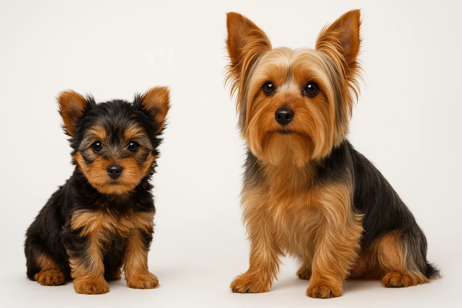
- Yorkshire Terrier — possibly due to coat characteristics that favor fungal colonization
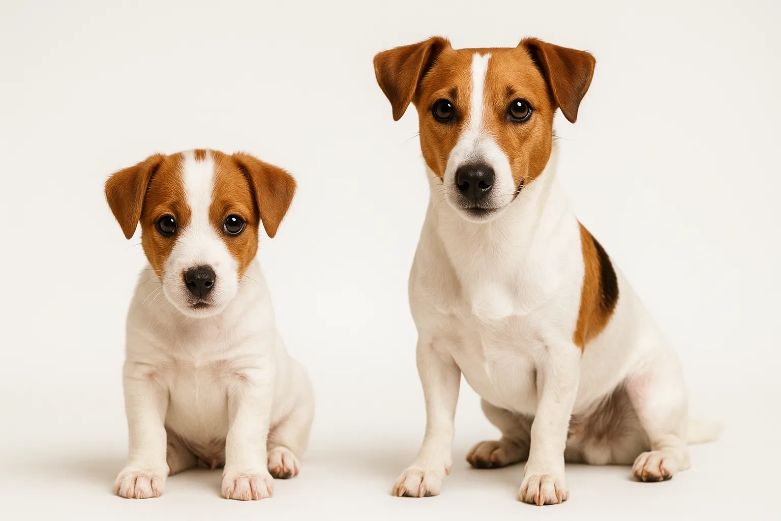
- Jack Russell Terrier — terrier breeds with earth-working heritage have higher exposure to soil-dwelling dermatophytes
- Persian cats (a key consideration in multi-species households, as cats are the most common reservoir for M. canis)
Risk is driven more by immune status and exposure than by breed genetics:
- Puppies under 12 months with immature immune systems
- Immunocompromised dogs on corticosteroids, chemotherapy, or with underlying illness
- Dogs in dense housing (shelters, breeding facilities, boarding kennels)
- Dogs with skin barrier compromise from other conditions like atopic dermatitis or skin allergies
- Hunting and working dogs with frequent soil and wildlife contact
How Ringworm Spreads
Transmission occurs through three primary routes:
Direct contact: Touching an infected animal. Cats are the primary reservoir for M. canis and can carry the fungus asymptomatically. Direct dog-to-dog transmission is common in shelters and kennels.
Contaminated environment: Fungal spores shed from infected hair and skin scales persist on surfaces, bedding, collars, brushes, and crates. A single infected dog can contaminate an entire household environment.
Soil contact: M. gypseum lives in soil, particularly soil enriched with keratin (from decomposing hair, feathers, or hooves). Digging dogs are at higher risk for this species.
The incubation period from exposure to visible lesions is typically 1-3 weeks but can be longer in dogs that mount a partial immune response.
Diagnosis
Accurate diagnosis is essential because ringworm mimics several other skin conditions, and treatment is pointless if the diagnosis is wrong.
Wood’s Lamp Examination
A Wood’s lamp emits ultraviolet light. Approximately 50% of M. canis strains produce a metabolite that fluoresces apple-green under UV light. This is a useful screening tool but has significant limitations: negative fluorescence does not rule out ringworm (half of M. canis strains and all other dermatophyte species do not fluoresce), and false positives occur from topical medications, lint, and dead skin scales.
Fungal Culture
The gold standard for ringworm diagnosis. Hair and scale samples from the lesion margins are placed on dermatophyte test medium (DTM) or Sabouraud dextrose agar. Cultures take 7-21 days to grow, and species identification confirms both the diagnosis and the likely source of infection. The delay is frustrating, but culture accuracy is far superior to other methods.
PCR Testing
Polymerase chain reaction testing for dermatophyte DNA provides faster results (24-48 hours) than culture and is highly sensitive. PCR is increasingly available and useful for both diagnosis and monitoring treatment response.
Trichography
Microscopic examination of plucked hairs can reveal fungal spores (arthroconidia) surrounding or within the hair shaft. This provides a rapid presumptive diagnosis but requires experience to interpret.
Rule Out Similar Conditions
Ringworm can be confused with bacterial folliculitis, mange, hormonal alopecia, sebaceous adenitis, and localized skin allergies. A fungal culture or PCR test prevents misdiagnosis and inappropriate treatment.
Treatment Protocols
Treatment of ringworm has two equally important goals: clearing the infection from the dog and decontaminating the environment. Neglecting either goal leads to reinfection.
Topical Therapy
Topical treatment reduces environmental contamination by killing fungal spores on the coat surface:
- Lime sulfur dips: Applied as a whole-body dip every 5-7 days. Effective and well-studied but has a strong sulfur odor, stains light-colored coats yellow, and irritates eyes and mucous membranes. Despite these drawbacks, lime sulfur remains one of the most reliable topical treatments.
- Miconazole/chlorhexidine shampoo: Combination antifungal/antiseptic shampoo used 2-3 times weekly. Less unpleasant than lime sulfur and effective for reducing spore shedding.
- Enilconazole: A topical antifungal rinse used in some countries but not widely available in the United States.
- Clotrimazole or miconazole cream: Useful for small, localized lesions as adjunctive therapy but insufficient as sole treatment for most cases.
Systemic Therapy
Oral antifungal medication is required for generalized infections, nail involvement, or cases that fail to respond to topical treatment alone:
- Itraconazole: First-line systemic antifungal for canine dermatophytosis. Typically dosed at 5 mg/kg once daily. Pulse therapy protocols (one week on, one week off) are effective and reduce cost and potential side effects. Well-absorbed with food, especially fatty meals.
- Terbinafine: An alternative to itraconazole with good efficacy. Dosed at 30 mg/kg once daily. Can be used in pulse protocols.
- Fluconazole: Sometimes used but generally considered less effective against dermatophytes than itraconazole or terbinafine.
- Griseofulvin: An older antifungal that was the historical standard of care. Still effective but has more potential side effects (bone marrow suppression, teratogenicity) than newer options and is used less frequently.
Treatment typically continues for a minimum of 6-8 weeks and should not be stopped based on visible lesion resolution alone. Two consecutive negative fungal cultures, taken 2-3 weeks apart, confirm mycological cure. Stopping treatment too early is the most common cause of relapse.
Environmental Decontamination
This step is non-negotiable. Without environmental cleanup, reinfection is virtually guaranteed.
- Vacuum all carpets, upholstered furniture, and fabric surfaces at least twice weekly. Dispose of vacuum bags or clean canisters after each use.
- Wash all bedding, blankets, collars, leashes, and washable toys in hot water with bleach (dilute household bleach at 1:10 concentration is effective against dermatophyte spores).
- Hard surfaces: Clean with dilute bleach or an accelerated hydrogen peroxide disinfectant.
- Discard items that cannot be adequately cleaned (cardboard scratching posts, heavily contaminated fabric items).
- Restrict the infected dog to easily cleaned rooms during treatment.
- Grooming tools: Soak in dilute bleach for 10 minutes or discard and replace.
Prevention Strategies
- Quarantine new animals entering the household for 2-3 weeks and screen for ringworm before introduction
- Maintain good hygiene in boarding and grooming facilities
- Avoid sharing grooming tools between dogs
- Test cats in the household if a dog develops ringworm (M. canis cross-species transmission is common)
- Support immune health through proper nutrition, parasite control, and stress reduction
- Minimize soil contact for susceptible dogs if M. gypseum is the identified species
- Clean and disinfect kennels, crates, and bedding regularly
Zoonotic Risk
Ringworm is one of the most commonly transmitted infections from animals to humans. Children, elderly individuals, and immunocompromised people are at highest risk. Human ringworm from canine/feline sources typically appears as itchy, circular, red patches on the skin.
If your dog is diagnosed with ringworm:
- Wash hands thoroughly after handling the dog
- Wear gloves when applying topical treatments
- Keep the dog off furniture and out of bedrooms if household members are immunocompromised
- Seek medical evaluation for any household member who develops suspicious skin lesions
Related Condition Pathways
Related Breed Longevity Guides
When to Seek Veterinary Care
Routine evaluation is appropriate for:
- Small, localized patches of hair loss without other signs
- Monitoring treatment response at scheduled intervals
Urgent evaluation is needed for:
- Rapidly spreading hair loss across multiple body regions
- Secondary bacterial infection (pustules, pain, swelling, foul odor)
- Ringworm in a puppy under 8 weeks, immunocompromised dog, or multi-pet household
- Household members developing skin lesions after dog diagnosis
Ringworm is rarely dangerous, but its contagious nature makes prompt, thorough treatment important for the entire household.
Frequently Asked Questions
How did my dog get ringworm? Your dog likely contracted ringworm through direct contact with an infected animal (including cats, which are the most common reservoir for M. canis), from a contaminated environment (grooming facility, boarding kennel, dog park), or from soil contact. The incubation period is 1-3 weeks, so exposure may have occurred well before lesions appeared.
Is ringworm dangerous to my dog? In otherwise healthy dogs, ringworm is a self-limiting infection that resolves even without treatment, though this can take several months. It is not life-threatening. The primary reasons to treat are to reduce the duration of infection, minimize environmental contamination, and prevent transmission to other animals and humans.
How long is my dog contagious? An infected dog sheds spores from the time lesions appear (and potentially before) until treatment achieves mycological cure, confirmed by negative fungal cultures. This typically takes 6-12 weeks with appropriate treatment. During this period, environmental decontamination measures should be maintained.
Can ringworm come back? Yes. Reinfection can occur from contaminated environments, contact with carrier animals (especially cats), or continued soil exposure. Dogs do develop some immunity after infection, but it is not complete, particularly in immunocompromised individuals. Thorough environmental decontamination during treatment reduces reinfection risk.
Do I need to shave my dog for treatment? Clipping hair around lesions and, in severe cases, full-body clipping can improve topical treatment penetration and reduce environmental spore shedding. However, clipping can also spread spores if not done carefully. Discuss the benefits and risks with your veterinarian based on the extent of your dog’s infection.
Can I still walk my dog during ringworm treatment? Yes, but avoid close contact with other dogs, and do not allow your dog to use communal grooming facilities or daycare during treatment. Leash walks on non-contact routes are fine. The primary transmission risk is through direct contact and environmental contamination, not brief outdoor proximity.
Medical Disclaimer
This guide is informational and does not replace in-person veterinary diagnosis or treatment. Ringworm requires accurate laboratory diagnosis (fungal culture or PCR), as many skin conditions look similar but require different treatment approaches. If multiple animals or household members are affected, coordinate care with both your veterinarian and your physician.
References
[1] Moriello KA, et al. “Diagnosis and treatment of dermatophytosis in dogs and cats: Clinical Consensus Guidelines of the World Association for Veterinary Dermatology.” Vet Dermatol. 2017;28(3):266-e68. [2] DeBoer DJ, Moriello KA. “Humoral and cellular immune responses to Microsporum canis in naturally and experimentally infected cats.” J Med Vet Mycol. 1993;31(2):121-132. [3] Moriello KA. “Feline dermatophytosis: aspects pertinent to disease management in single and multiple cat situations.” J Feline Med Surg. 2014;16(5):419-431. [4] Cafarchia C, et al. “Isolation of Microsporum canis from the hair coat of pet dogs and cats belonging to owners diagnosed with M. canis tinea corporis.” Vet Dermatol. 2006;17(5):327-331. [5] Scott DW, Miller WH, Griffin CE. Muller & Kirk’s Small Animal Dermatology. 7th ed. Elsevier; 2013.