Evidence deep dives for Atlantoaxial Instability
Pair mechanism-level evidence with practical protocol context before discussing next steps with your veterinarian.
A Tiny Joint with Life-Altering Consequences
Atlantoaxial instability (AAI) strikes the junction between the first two vertebrae in the neck — the atlas (C1) and the axis (C2). In a healthy dog, this joint allows controlled head movement while a bony pivot pin called the dens and a complex of ligaments protect the spinal cord. In toy and small-breed dogs, the dens may be absent, underdeveloped, or inadequately anchored. Without that structural anchor, the atlas can shift and compress the spinal cord.
The consequences range from neck pain to sudden, complete paralysis of all four limbs.
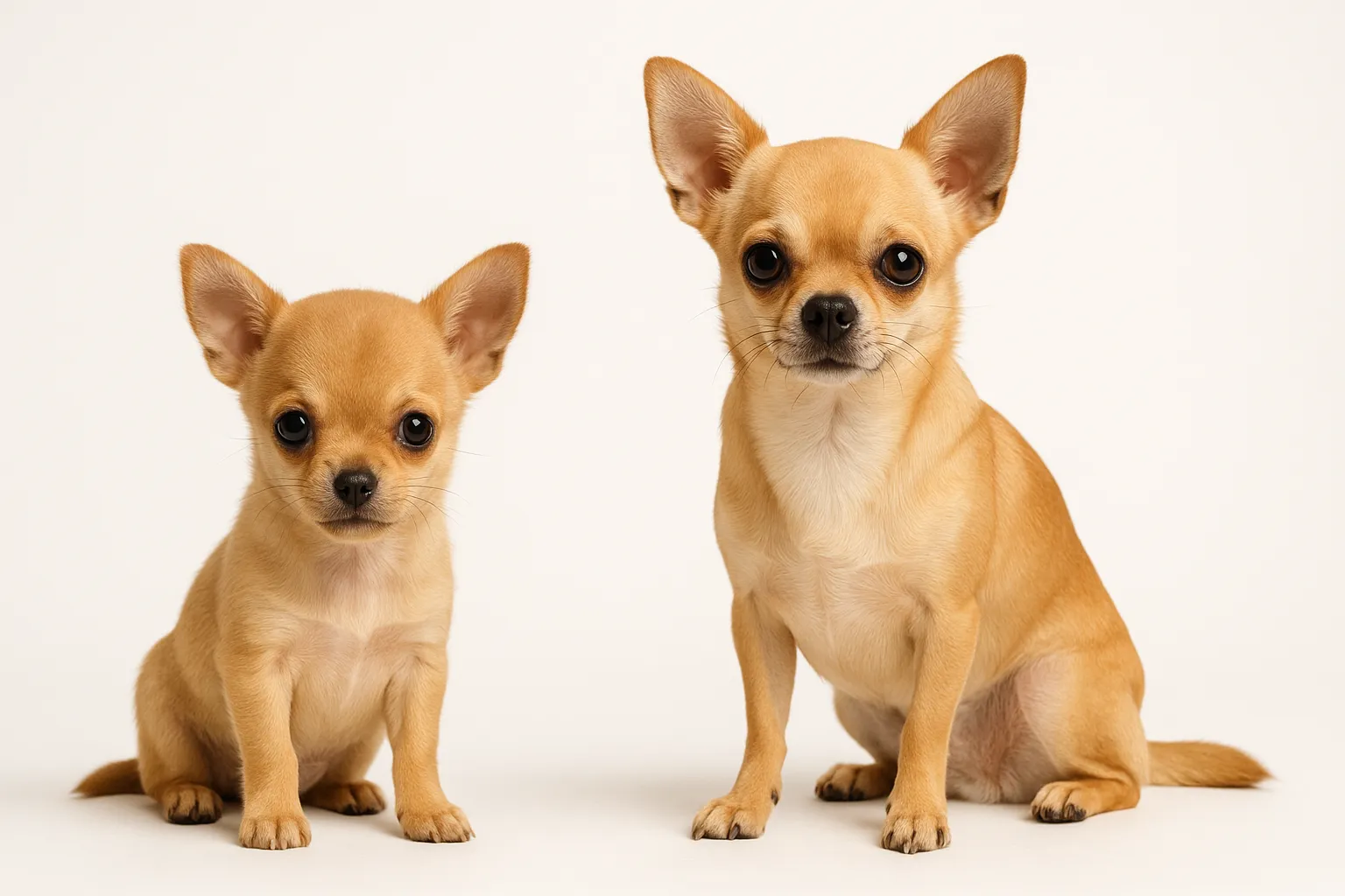
This condition overwhelmingly affects toy and miniature breeds. Their miniaturized skeletal structures make even modest spinal cord compression a proportionally larger neurological insult. A Chihuahua with AAI faces a fundamentally different risk profile than a Labrador with the same degree of joint movement.
A less common form occurs when trauma — sometimes as minor as jumping off a couch or being accidentally stepped on — ruptures the ligament holding the dens in position. This can happen in small dogs without any congenital abnormality.
Why This Demands Urgent Attention
The spinal cord at the cranial cervical level controls all four limbs, respiration, and numerous vital functions. A dog can progress from neck pain to quadriplegia with a single wrong movement. Without surgical stabilization, the risk of acute deterioration hangs over every routine activity.
From a longevity perspective, repeated spinal cord compression causes cumulative, potentially permanent damage. Dogs treated with surgical fusion early — before irreversible cord injury — have the best long-term neurological outcomes. Waiting until paralysis occurs dramatically worsens the prognosis.
Recognizing the Warning Signs
AAI presents across a spectrum of severity. If your small-breed dog shows any of these signs, especially after minor trauma, seek evaluation immediately.
Early or mild signs:
- Crying out when picked up, resisting head movement, or holding the neck stiffly
- Reluctance to lower the head to eat or drink from floor-level bowls
- Forelimb weakness or stumbling, particularly after jumping or playing
- A characteristic “praying position” — front legs extended with elbows on the floor, hindquarters elevated — as the dog tries to take weight off the painful neck
- Knuckling the paws or stumbling on all four limbs (proprioceptive deficits)
Acute crisis signs:
- Sudden collapse after minor trauma
- Rapid progressive weakness in all four limbs
- Complete inability to move (quadriplegia)
Any small-breed dog with acute neck pain after minor trauma, or a young toy-breed dog with progressive wobbliness, warrants emergency evaluation. This is not a condition to manage with rest and anti-inflammatories alone.
How AAI Is Diagnosed
Advanced imaging is required. Plain X-rays of the cervical spine can suggest subluxation (widened C1-C2 space, absent or malformed dens), but sensitivity is limited.
A critical safety note: flexed-neck X-ray views to demonstrate instability should never be performed in suspected AAI. The positioning can cause acute cord compression and neurological catastrophe.
MRI of the cervical spine is the gold standard. It directly visualizes spinal cord compression, cord signal changes indicating injury, and soft tissue structures.
CT provides superior bone detail for surgical planning, particularly for evaluating dens morphology and identifying screw placement points.
Both modalities require general anesthesia with meticulous patient positioning to avoid worsening neurological status during the procedure.
Complete diagnostic workup:
- MRI of the cervical spine for cord compression and injury assessment
- CT scan for dens evaluation and surgical planning
- Lateral cervical radiography (neutral position only, never flexion views) for initial assessment in stable patients
- Full neurological examination to grade severity
- Baseline CBC and chemistry panel for anesthetic risk assessment
Surgical Stabilization vs. Conservative Management
Surgery (atlantoaxial fusion) is the recommended definitive treatment for dogs with moderate to severe neurological deficits or recurrent neck pain. The procedure fuses C1 and C2 using bone screws, pins, and polymethylmethacrylate, eliminating the abnormal motion that threatens the cord. In experienced hands, outcomes are good to excellent in 70-90% of cases when performed before irreversible cord damage.
Conservative management — cervical splints, neck braces, strict activity restriction, and anti-inflammatory medications — is appropriate for dogs with mild signs or as a bridge to surgery. But it carries significant failure rates. Dogs managed conservatively remain at ongoing risk of acute decompensation from everyday events. Conservative care buys time; it does not fix the underlying instability.
Regardless of treatment approach, immediate safety measures include:
- Replace neck collars with harnesses immediately (collars apply force directly at the C1-C2 level)
- Strict activity restriction: no jumping, no rough play, no sudden head movements
- Ramps or carrying for all elevated surface access
- Slightly elevated food and water bowls to reduce cervical flexion
- Anti-inflammatory medications only as a bridge, not as a substitute for surgical evaluation
Refer to a veterinary neurologist for MRI/CT and surgical consultation as soon as AAI is suspected.
12-Week Post-Diagnosis Execution Plan
- Weeks 1-2 (baseline lock-in): Confirm the diagnosis, start a shared household log, and track daily markers including neck posture, limb function, appetite, and sleep quality.
- Weeks 3-4 (adherence audit): Verify every caregiver follows the same activity restrictions. Identify where the protocol breaks down (visitors picking up the dog incorrectly, unsupervised furniture jumping) and close the gap.
- Weeks 5-6 (response checkpoint): Compare neurological function against baseline. If gait, strength, or head position is worsening, escalate immediately rather than waiting for the scheduled recheck.
- Weeks 7-8 (risk tightening): Define exact thresholds for emergency contact. Every caregiver should understand that sudden inability to walk, acute neck pain with crying, or loss of bladder control means go now.
- Weeks 9-10 (resilience build): Reinforce the safe mobility routines your neurologist has cleared. Ensure activity restrictions have become permanent household habits.
- Weeks 11-12 (handoff to maintenance): Document the long-term monitoring cadence, confirm which weekly metrics to continue tracking, and schedule the next neurological recheck.
The Pattern That Leads to Preventable Crises
Families tend to react only to dramatic events, but AAI outcomes improve when response starts at the first measurable change. Missing a narrow window for reassessment can turn a manageable situation into permanent neurological damage.
Common process failures:
- Inconsistent household execution. Different caregivers apply different rules about carrying, collar use, or furniture access.
- Over-correcting too fast. Changing medications, activity level, and feeding setup simultaneously obscures what is helping.
- Waiting for the dramatic event. Teams that review one objective metric weekly (gait quality, head position, stumbling frequency) catch deterioration much earlier than those who wait for a crisis.
Nutrition and Recovery Support
Maintaining lean body condition is especially important. Excess weight increases the load on an already unstable cervical joint and worsens clinical signs.
Dogs recovering from fusion surgery need adequate protein for tissue healing. Anti-inflammatory omega-3 fatty acids at evidence-based doses may provide modest adjunctive benefit during recovery, though direct evidence in AAI recovery is absent.
- Feeding Guide for Toy Breeds
- Omega-3 Fish Oil for Dogs: Evidence and Dosing
- Bone Broth for Dogs provides a palatable, nutrient-dense liquid option during post-surgical recovery when appetite may be reduced
For evidence context and execution details, review:
- Muscle and Mobility Longevity Protocol
- Pain Assessment in Senior Dogs
- Canine Size-Lifespan Tradeoffs by Breed
What Monitoring Looks Like After Diagnosis
Monitoring tracks both disease stability and surgical recovery:
- Weekly home neurological assessments: Is gait improving, stable, or worsening? Is head position normal?
- Post-surgical rechecks at 2 weeks (incision check), 6 weeks (early neurological reassessment), and 3 months (imaging confirmation of fusion)
- Watch for implant failure or migration in surgically treated dogs — report any sudden neurological worsening immediately
- Monthly weight checks in conservatively managed dogs, because any weight gain worsens joint stress
Dogs with successful surgical fusion and preserved pre-operative cord function can have excellent long-term quality of life. Dogs that were quadriplegic before surgery have more variable outcomes depending on the duration and severity of cord compression.
When to Seek Emergency Care Immediately
These are neurological emergencies:
- Sudden inability to use all four limbs or sudden collapse
- Sudden cry of pain followed by inability to raise the head or move the neck
- Acute loss of bladder or bowel control
- Labored or irregular breathing in a dog with known AAI (suggests high cervical cord compromise affecting respiratory centers)
- Any rapid neurological deterioration in a conservatively managed dog
Related Condition Pathways
AAI shares clinical features and sometimes coexists with other spinal conditions in small breeds:
- Intervertebral Disc Disease: both cause spinal cord compression in small breeds; imaging distinguishes them.
- Spinal Disorders: cervical malformation and concurrent spinal instability may overlap in susceptible breeds.
- Arthritis: chronic cervical instability promotes degenerative joint changes that compound pain and mobility limitation over time.
Use these pages to build understanding and inform your conversations with your vet. Treatment decisions should always be confirmed clinically.
Related Breed Longevity Guides
AAI is overwhelmingly a toy and miniature breed condition:
Chihuahuas and Yorkshire Terriers have the highest reported prevalence. If you own a toy or miniature breed and notice sudden neck pain or acute weakness, consider AAI immediately.
Frequently Asked Questions
Can my dog recover from atlantoaxial instability without surgery?
Some dogs with mild signs can be managed conservatively with splints, braces, strict rest, and medications. But conservative management has significant failure rates and does not address the underlying mechanical instability. Most conservatively managed dogs remain at ongoing risk for sudden, severe deterioration. Surgical stabilization provides the most durable protection against progressive cord injury.
How long is recovery after atlantoaxial fusion surgery?
Initial hospitalization typically lasts 2-5 days. Activity restriction continues for 6-12 weeks while bone fusion consolidates. Full neurological improvement, when achievable, may continue for 6-12 months after surgery. Dogs with mild to moderate deficits before surgery typically show progressive improvement. Dogs that were quadriplegic pre-operatively have variable outcomes.
Why does my dog need a harness instead of a collar?
Neck collars apply traction directly at the C1-C2 joint level. In a dog with AAI, any force through the collar — leash pulls, sudden movements — can directly worsen subluxation and compress the cord. Harnesses distribute force across the chest, completely bypassing the vulnerable cervical spine. All dogs with AAI should use harnesses permanently.
Is atlantoaxial instability hereditary?
The congenital form involving dens aplasia or hypoplasia is considered heritable in toy breeds, though the exact genetic mechanisms are not fully mapped. Affected dogs should not be bred. The traumatic form caused by ligament rupture is not hereditary.
What is the prognosis for a dog with atlantoaxial instability?
Prognosis depends heavily on neurological status before treatment. Dogs with neck pain only (no motor deficits) treated surgically have excellent outcomes. Dogs with mild to moderate motor deficits treated surgically recover well in 70-85% of cases. Dogs with acute quadriplegia have guarded prognoses — outcomes vary with cord injury severity and timing of surgery.
Medical Disclaimer
This content is for educational purposes only and does not constitute veterinary medical advice. Atlantoaxial instability is a serious neurological condition requiring urgent veterinary evaluation including advanced imaging (MRI/CT). Treatment decisions, particularly regarding surgical versus conservative management, should be made with a veterinary neurologist or neurosurgeon.
References
- Platt SR, Chambers JN, Cross A. A modified ventral fixation for surgical management of atlantoaxial subluxation in 19 dogs. Vet Surg. 2004;33(4):349-354.
- Beaver DP, Ellison GW, Lewis DD, et al. Risk factors affecting the outcome of surgery for atlantoaxial subluxation in dogs: 46 cases (1978-1998). J Am Vet Med Assoc. 2000;216(7):1104-1109.
- Havig ME, Cornell KK, Hawthorne JC, et al. Evaluation of nonsurgical treatment of atlantoaxial subluxation in dogs: 19 cases (1992-2001). J Am Vet Med Assoc. 2005;227(2):257-262.
- Dube A, Moreau M, d’Anjou MA, et al. Atlantoaxial subluxation in miniature and toy breeds of dogs: a review of 21 cases. Can Vet J. 2008;49(12):1195-1200.
- Sanders SG, Bagley RS, Silver GM, et al. Outcomes and complications associated with ventral screws, pins, and polymethyl methacrylate for atlantoaxial instability in 12 dogs. J Am Anim Hosp Assoc. 2004;40(3):204-210.
Related reads
Related Reading
Continue exploring