Evidence deep dives for Degenerative Lumbosacral Stenosis
Pair mechanism-level evidence with practical protocol context before discussing next steps with your veterinarian.
The Tail Stops Wagging Before the Legs Give Out
It starts with something subtle — the tail that used to beat against your leg at the door now hangs limp, barely moving. Then the hind legs grow slow on stairs. Then one morning, your dog cannot rise from the floor without help. By the time most owners connect these dots, degenerative lumbosacral stenosis has been compressing nerve roots for months.
The lumbosacral junction — where the last lumbar vertebra (L7) meets the sacrum (S1) — bears enormous mechanical stress with every step, every jump, every excited sprint across the yard. Over time, disc degeneration, ligament thickening, and bone changes narrow the spinal canal at this critical point, squeezing the cauda equina: the nerve bundle that controls the hind limbs, tail, bladder, and anal sphincter.
The condition is sometimes called cauda equina syndrome, though that term more accurately describes the clinical presentation than the underlying degenerative process. DLS is the structural disease; cauda equina compression is the functional consequence.
Long-Term Consequences and Prevention Value
DLS directly threatens a dog’s quality of life through a predictable cascade:
- chronic lumbosacral pain that worsens with activity and position changes
- progressive hind-limb weakness and exercise intolerance
- bladder and bowel dysfunction in advanced cases
- secondary muscle atrophy from disuse and compensatory movement patterns
- progressive mobility loss that erodes independence and daily function
Because DLS develops gradually, many owners attribute early signs to “slowing down with age” rather than recognizing a treatable structural problem. Early identification and intervention can substantially slow functional decline and preserve comfort for years.
Which Dogs Are at Risk
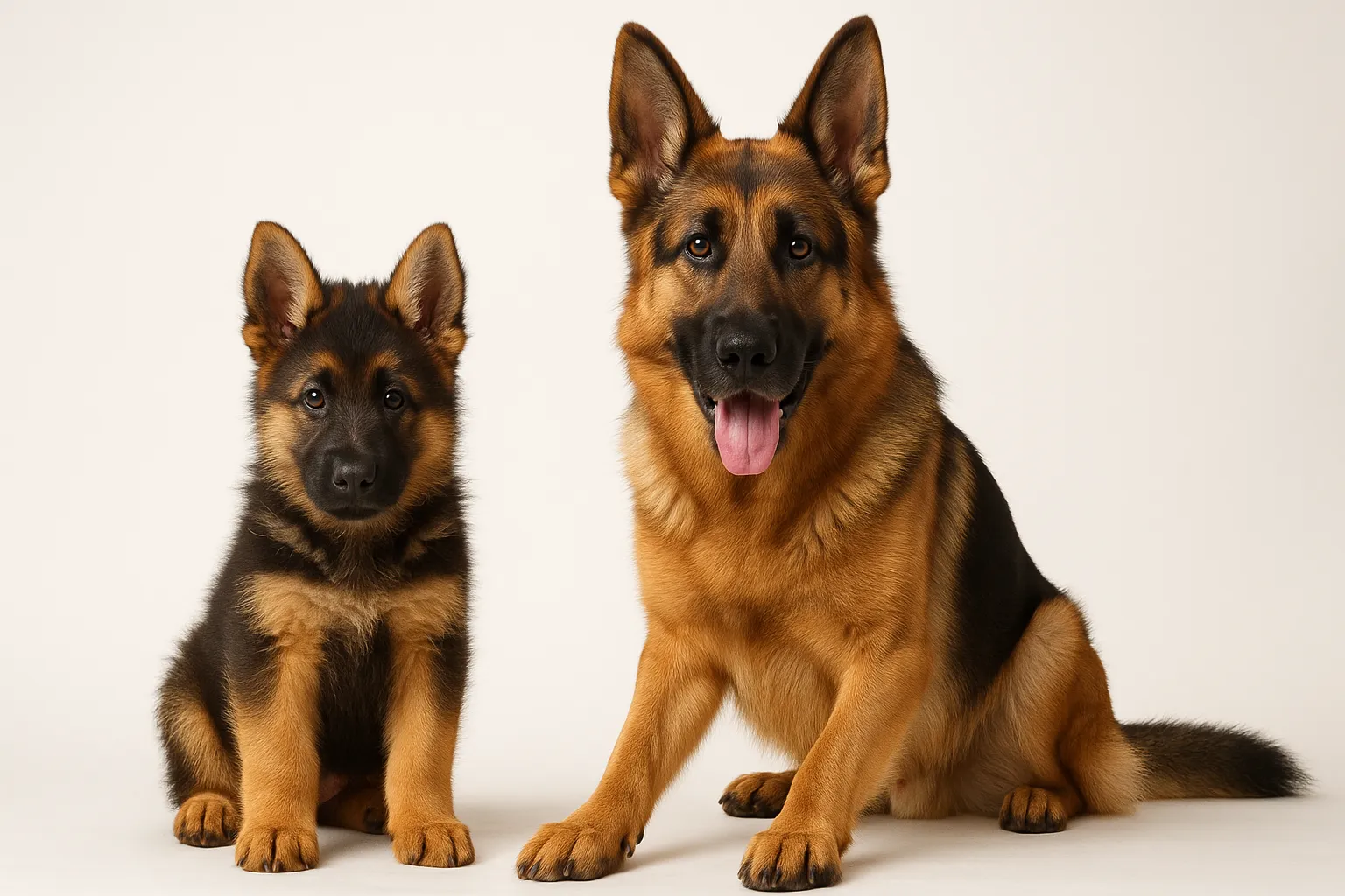
Large-breed dogs are disproportionately affected, with German Shepherds representing the single most overrepresented breed in clinical studies. The lumbosacral angle in German Shepherds may predispose them to greater mechanical stress at L7-S1 compared to other breeds.
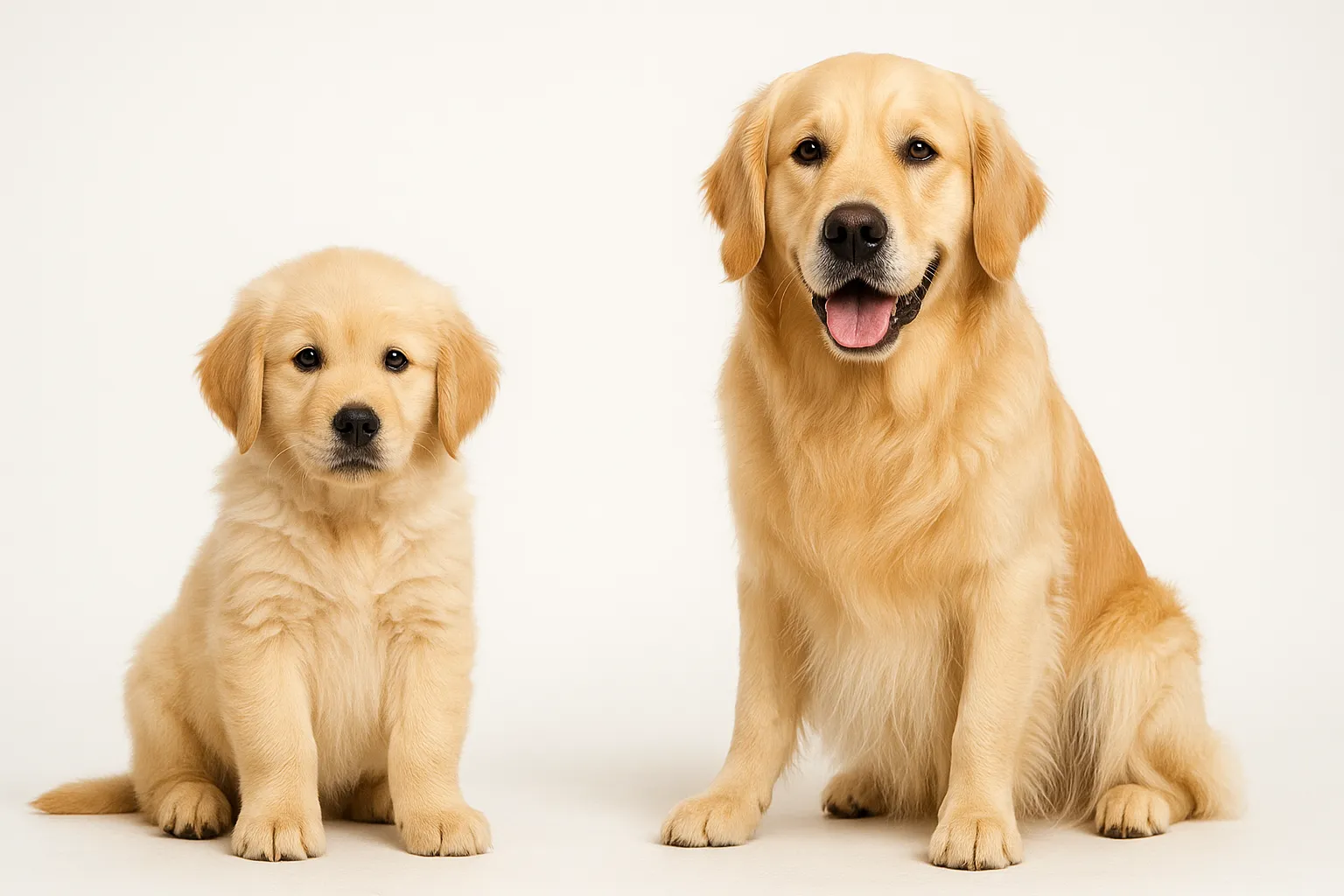
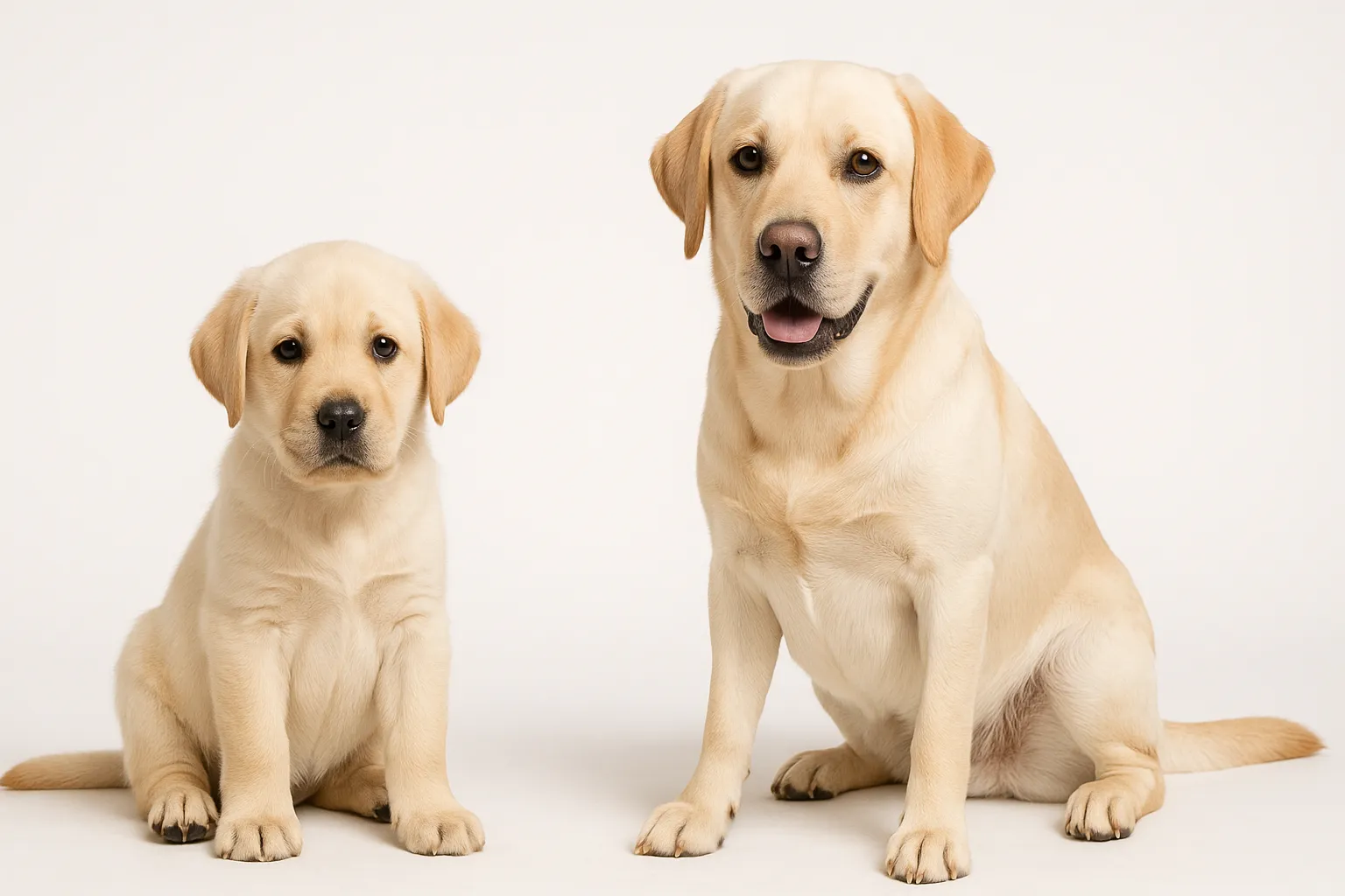
Other commonly affected breeds include Labrador Retrievers, Golden Retrievers, Rottweilers, Siberian Huskies, and Border Collies. Working and sporting dogs face additional risk from repetitive high-impact activities — jumping, agility, Schutzhund, and sustained running — that increase cumulative load on the lumbosacral junction.
Male dogs appear to be affected more frequently than females, though the sex distribution data is influenced by referral patterns. Overweight dogs of any breed experience accelerated degeneration due to increased mechanical loading.
Recognizing the Early Signs
The clinical presentation of DLS depends on which nerve roots are most compressed and how severely. Early signs are often subtle:
- reluctance to jump, climb stairs, or rise from a lying position
- pain on palpation of the lumbosacral region (the dog may flinch, vocalize, or sit down when pressure is applied)
- low tail carriage or decreased tail wagging
- intermittent hind-limb lameness that does not localize to a single joint
- exercise intolerance — the dog starts strong but weakens or slows after 10-20 minutes
- shifting weight forward to unload the hindquarters
- difficulty posturing to urinate or defecate
As the disease progresses, more definitive neurologic signs emerge:
- proprioceptive deficits in the hind limbs (knuckling, scuffing toenails)
- muscle atrophy in the hindquarters and pelvic limb musculature
- urinary or fecal incontinence (indicates significant nerve compression)
- decreased anal tone on examination
- progressive paraparesis (hind-limb weakness)
A key differentiator from hip dysplasia — which produces similar hind-end reluctance — is the pain response to direct lumbosacral palpation and the tail-related changes. Dogs with DLS typically show pain when the lumbosacral junction is pressed dorsoventrally, and tail dysfunction is uncommon in isolated hip disease.
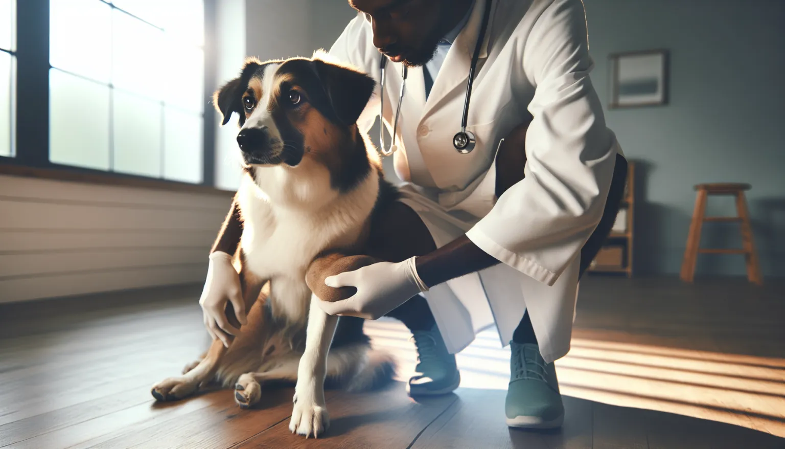
What Testing and Evaluation Looks Like
Physical and Neurologic Examination
A thorough neurologic examination is the foundation. The veterinarian will assess:
- gait and posture symmetry
- proprioception in all four limbs (hind limbs specifically)
- spinal reflexes — patellar, withdrawal, perineal
- pain response to lumbosacral extension (lordosis test): extending the hips while applying dorsal pressure over L7-S1 typically reproduces pain in affected dogs
- rectal examination to assess anal tone and detect pain on pelvic floor palpation
- tail tone and voluntary tail movement
Advanced Imaging
Radiographs may show spondylosis deformans (bony bridging), disc space narrowing, and endplate sclerosis at L7-S1, but these changes do not correlate reliably with clinical severity. Many older large-breed dogs have radiographic lumbosacral changes without clinical signs.
MRI is the gold standard for evaluating DLS. It reveals:
- disc protrusion and degree of canal narrowing
- nerve root compression and displacement
- soft tissue contributions (ligament hypertrophy, epidural fibrosis)
- disc hydration status and degeneration grade
CT (with or without myelography) provides excellent bony detail and is useful for surgical planning, though MRI offers superior soft tissue resolution.
Electrodiagnostics (EMG and nerve conduction studies) can confirm denervation in the muscles supplied by the cauda equina and help localize the lesion.
Differential Diagnosis
Several conditions mimic DLS and must be excluded or identified as concurrent problems:
- hip dysplasia — often coexists, especially in German Shepherds
- intervertebral disc disease — can occur at other spinal levels
- degenerative myelopathy — progressive but non-painful, unlike DLS
- lumbosacral neoplasia — tumors at L7-S1
- discospondylitis — infection of the disc and adjacent vertebrae
- peripheral neuropathy
The combination of lumbosacral pain plus neurologic deficits plus breed predisposition creates a strong index of suspicion, but imaging confirmation is essential before committing to invasive treatment.
Conservative Management
Many dogs with mild to moderate DLS respond well to multimodal conservative management, particularly when started early.
Weight Management
Excess body weight directly increases mechanical load on the lumbosacral junction. Achieving and maintaining a body condition score of 4-5/9 is one of the single highest-impact interventions. Even a 10% reduction in body weight can meaningfully reduce pain and improve function.
Activity Modification
The goal is to maintain muscle mass and cardiovascular fitness while avoiding activities that stress the lumbosacral junction:
- eliminate jumping, rough play, and high-impact landing
- replace free running with controlled leash walks on even terrain
- swimming and underwater treadmill provide excellent low-impact exercise
- avoid prolonged sitting or lying in positions that flex the lumbosacral spine
- short, frequent exercise sessions rather than long, exhausting ones
Pain Management
Multimodal analgesia typically includes:
- NSAIDs as the first-line anti-inflammatory (with appropriate GI and renal monitoring)
- gabapentin or pregabalin for neuropathic pain
- tramadol as an adjunctive analgesic in some protocols
- muscle relaxants when secondary muscle spasm contributes to pain
Physical Rehabilitation
Structured rehabilitation programs improve outcomes:
- core strengthening exercises to stabilize the lumbosacral region
- range-of-motion exercises for the hind limbs
- proprioceptive training (balance boards, cavaletti rails)
- therapeutic ultrasound and laser therapy for local pain relief
- hydrotherapy — swimming and underwater treadmill
A veterinary rehabilitation specialist can design a program matched to the dog’s current function level and adjust it as the condition evolves.
Supplements
Evidence for supplements in DLS specifically is limited, but several have biological plausibility for supporting nerve and joint health:
- omega-3 fatty acids — anti-inflammatory properties that may reduce nerve root inflammation
- glucosamine and chondroitin — may support cartilage health in facet joints
- SAMe (S-adenosylmethionine) — neuroprotective properties in some models
- vitamin E — antioxidant support for nerve tissue
Supplements are adjunctive, not primary therapy. They do not reverse structural narrowing.
Surgical Options
Surgery is considered when conservative management fails to control pain or when neurologic deficits are progressing.
Dorsal Laminectomy
The most common surgical approach involves removing the dorsal lamina of L7 and sometimes S1 to decompress the cauda equina. This directly increases canal space and relieves pressure on the nerve roots.
Reported success rates range from 50-93% in the literature, depending on case selection, severity at the time of surgery, and the specific technique used. Dogs with pain as the primary complaint tend to respond better than those with advanced neurologic deficits.
Foraminotomy
When nerve root compression occurs primarily in the lateral foramina (the exit channels), targeted foraminotomy can decompress specific nerve roots without destabilizing the entire segment.
Stabilization Procedures
Some surgeons combine decompression with lumbosacral stabilization (fixation with screws, pins, or PMMA) when instability contributes to the compression. Stabilization prevents ongoing dynamic compression but adds surgical complexity and complication risk.
Post-Surgical Outcomes
Most dogs show meaningful improvement in pain and function within 4-8 weeks of surgery. Full recovery may take 3-6 months with structured rehabilitation. Surgical complications include infection, implant failure (in stabilization cases), and recurrence of compression from scar tissue formation.
Dogs with urinary or fecal incontinence before surgery have a less predictable recovery — some regain continence, but those with long-standing nerve damage may not.
Long-Term Monitoring
DLS is a progressive condition. Even with successful conservative or surgical management, ongoing monitoring is essential:
- neurologic reassessment every 3-6 months
- track gait quality, exercise tolerance, and proprioceptive function
- monitor for urinary or fecal changes
- reassess body condition and muscle mass trends
- adjust activity protocols and pain management as the condition evolves
Owners should maintain a simple weekly log of:
- willingness to rise, jump, or climb
- tail carriage and wagging frequency
- exercise duration before slowing or reluctance
- any episodes of hind-limb weakness, knuckling, or stumbling
- toileting posture and any incontinence episodes
Trend data is far more informative than single observations. Gradual decline is easy to miss without structured tracking.
When This Becomes an Emergency
Seek immediate veterinary care for:
- sudden onset of hind-limb paralysis or inability to stand
- acute loss of bladder or bowel control
- severe pain unresponsive to prescribed medication
- rapid deterioration in neurologic function over hours to days
Seek prompt same-day care for:
- new onset of urinary hesitation or incontinence
- progressive knuckling or dragging of hind feet
- marked increase in pain despite current management
- significant decline in exercise tolerance over 1-2 weeks
Preventing Accelerated Degeneration
While DLS cannot be fully prevented in predisposed breeds, several strategies may slow progression:
- maintain lean body condition throughout life
- avoid repetitive high-impact activities during skeletal maturation
- build and maintain strong core musculature through targeted exercise
- address early signs promptly rather than waiting for advanced disease
- screen working and sporting dogs proactively if breed and activity level create elevated risk
For German Shepherds and other high-risk breeds, discuss lumbosacral screening with your veterinarian by age 5-6, even in the absence of clinical signs. Early structural changes on imaging may justify preemptive activity modification.
Longevity Science Connections
- Hip Dysplasia
- Intervertebral Disc Disease (IVDD)
- Degenerative Myelopathy
- Arthritis
- Muscle and Mobility Longevity Protocol
- Senior Dog Screening Protocol
Frequently Asked Questions
What is the difference between degenerative lumbosacral stenosis and IVDD?
DLS specifically affects the L7-S1 junction and compresses the cauda equina nerve roots. IVDD can occur at any spinal level and compresses the spinal cord or nerve roots depending on location. The underlying degenerative mechanisms overlap, but the clinical presentations and surgical approaches differ.
Can degenerative lumbosacral stenosis be cured?
No. DLS is a progressive structural condition. Treatment aims to manage pain, preserve function, and slow progression. Surgery can decompress the nerve roots and provide significant relief, but the underlying degenerative process continues.
Is this the same as cauda equina syndrome?
DLS is the most common cause of cauda equina syndrome in dogs, but cauda equina syndrome is a clinical description (compression of the cauda equina nerve bundle), not a specific disease. Other causes include tumors, infections, and fractures.
How do I know if my dog’s hind-end weakness is from DLS or hip dysplasia?
Both conditions cause hind-end reluctance and exercise intolerance. Key differentiators include lumbosacral pain on palpation, tail dysfunction, and neurologic deficits (knuckling, proprioceptive loss) — which point toward DLS rather than isolated hip disease. Advanced imaging is typically required for definitive differentiation. Both conditions can coexist.
Will my dog need surgery?
Not necessarily. Many dogs respond well to conservative management, especially when started early. Surgery is reserved for dogs with progressive neurologic deficits or pain that does not respond adequately to medical management.
Can physical rehabilitation help?
Physical rehabilitation is one of the most effective conservative interventions. Core strengthening, hydrotherapy, and controlled exercise programs can improve function and slow decline. Rehabilitation is also critical after surgery.
Should I restrict my dog’s activity completely?
No. Complete rest leads to muscle atrophy and deconditioning, which worsens the condition. The goal is controlled, low-impact activity that maintains muscle mass without overloading the lumbosacral junction. Swimming and underwater treadmill work are particularly beneficial.
Medical Disclaimer
This article is educational and not a substitute for veterinary care. Dogs showing signs of hind-limb weakness, pain, or incontinence require professional evaluation and diagnosis.
Supporting Recovery and Prevention Through Diet
Degenerative Lumbosacral Stenosis management benefits from a feeding strategy that supports lean body condition and reduces systemic inflammation.
- Omega-3 Fish Oil for Dogs: anti-inflammatory support that may help manage nerve root inflammation.
- Glucosamine and Chondroitin for Dogs: may support cartilage health in degenerating facet joints.
- Feeding Guide for Senior Dogs: Healthspan Nutrition: structured nutritional support for aging dogs with chronic conditions.
Any protocol adjustment — timing, dose, or addition — should be confirmed with your veterinarian before implementation.
Related Condition Pathways
Degenerative lumbosacral stenosis frequently coexists with or mimics other conditions affecting the hind end. Understanding these overlaps improves diagnostic accuracy and treatment planning.
- Hip Dysplasia: Often coexists in large-breed dogs, making clinical differentiation challenging without advanced imaging.
- Arthritis: Facet joint arthropathy at L7-S1 is part of the DLS complex, and concurrent peripheral joint arthritis compounds mobility loss.
- Degenerative Myelopathy: Progressive hind-limb weakness without pain — key clinical distinction from DLS, which is typically painful.
- Intervertebral Disc Disease (IVDD): Can occur concurrently at other spinal levels, complicating the clinical picture.
- Obesity: Excess body weight directly accelerates lumbosacral degeneration and worsens nerve compression.
Related Breed Longevity Guides
Breed-specific risk context helps determine screening timing and management intensity for lumbosacral disease.
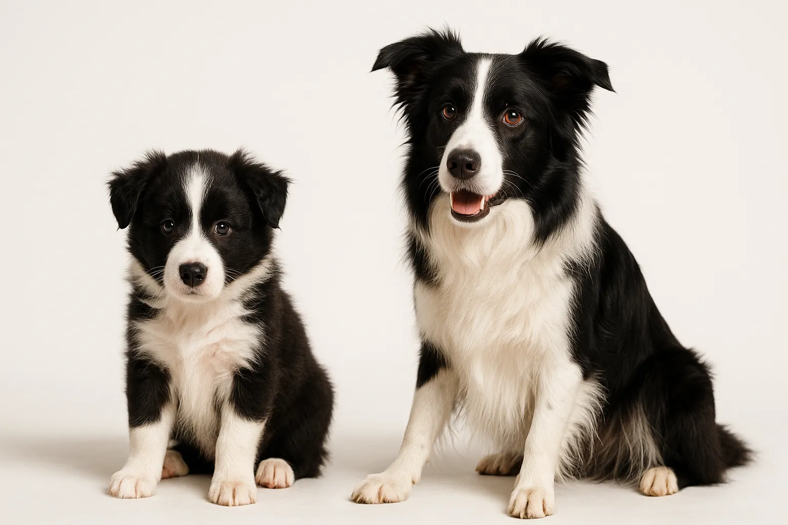
- German Shepherd Lifespan & Longevity Guide: the single most overrepresented breed, justifying proactive lumbosacral screening by age 5-6.
- Labrador Retriever Lifespan & Longevity Guide: high risk compounded by breed tendency toward obesity.
- Golden Retriever Lifespan & Longevity Guide: predisposition context supports lower thresholds for early investigation.
- Rottweiler Lifespan & Longevity Guide: large frame and working-dog activity patterns increase lumbosacral load.
- Siberian Husky Lifespan & Longevity Guide: sustained running and pulling activities create cumulative lumbosacral stress.
- Border Collie Lifespan & Longevity Guide: high-intensity activity patterns in this breed warrant attention to spinal health.
References
- Meij BP, Bergknut N. Degenerative lumbosacral stenosis in dogs. Vet Clin North Am Small Anim Pract. 2010;40(5):983-1009.
- De Risio L, et al. Association of clinical and magnetic resonance imaging findings with outcome in dogs with presumptive acute noncompressive nucleus pulposus extrusion. J Am Vet Med Assoc. 2009;234(4):495-504.
- Suwankong N, et al. Review on degenerative lumbosacral stenosis in dogs and assessment of diagnostic imaging. Vet Comp Orthop Traumatol. 2008;21(4):299-311.
- Worth AJ, et al. Computed tomographic evaluation of the lumbosacral junction in working dogs with and without clinical signs of cauda equina compression. Vet Radiol Ultrasound. 2009;50(3):306-313.
Related reads
Related Reading
Continue exploring