Evidence deep dives for Pemphigus
Pair mechanism-level evidence with practical protocol context before discussing next steps with your veterinarian.
When the Immune System Attacks the Glue That Holds Skin Together
It often starts quietly: a patch of crusty, scaly skin on the bridge of the nose. Thickened, cracked footpads. Crusty lesions on the ear flaps that do not respond to antibiotics. For weeks or months, pemphigus can be mistaken for allergies, infection, or just dry skin — until it spreads.
Pemphigus is a group of autoimmune skin diseases in which the immune system produces antibodies against desmoglein, the protein that holds skin cells together. When these intercellular bonds break (a process called acantholysis), fragile blisters form just beneath the skin surface. They rupture almost immediately, leaving behind the crusts, erosions, and ulcers that define the disease clinically.
Pemphigus foliaceus (PF) accounts for approximately 90% of canine pemphigus cases. It targets the superficial epidermis, and intact blisters are almost never seen — they are too fragile and too shallow. What owners and veterinarians see instead are pustules, thick crusts, scales, and raw erosions, typically beginning on the face, ears, and footpads before potentially spreading across the body.
Other variants exist but are rare: pemphigus vulgaris (deeper blistering, much more severe), pemphigus erythematosus (a milder, localized form often limited to the nose), and paraneoplastic pemphigus (driven by an underlying tumor).
Living With Pemphigus: The Longevity Equation
Pemphigus is a chronic condition. Most dogs require immunosuppressive therapy for life, and that creates a two-sided longevity equation.
The disease side: untreated PF can spread across large areas of the body, causing severe pain, protein loss through weeping lesions, and life-threatening secondary bacterial infections that penetrate the broken skin barrier.
The treatment side: the immunosuppressive drugs that control PF also reduce immune surveillance against infections, and long-term corticosteroid use brings its own metabolic costs — weight gain, muscle weakness, skin thinning, increased diabetes risk.
The balance point matters. Well-managed PF allows many dogs to live normal lifespans with reasonable quality of life. Poorly controlled disease, or disease that resists standard immunosuppression, carries higher mortality from both the disease itself and treatment complications.
Breeds Where Pemphigus Concentrates
Pemphigus foliaceus shows strong breed predisposition:
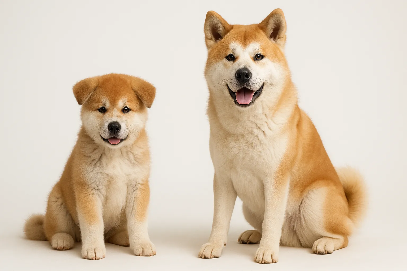
- Akita: one of the most over-represented breeds in PF registries
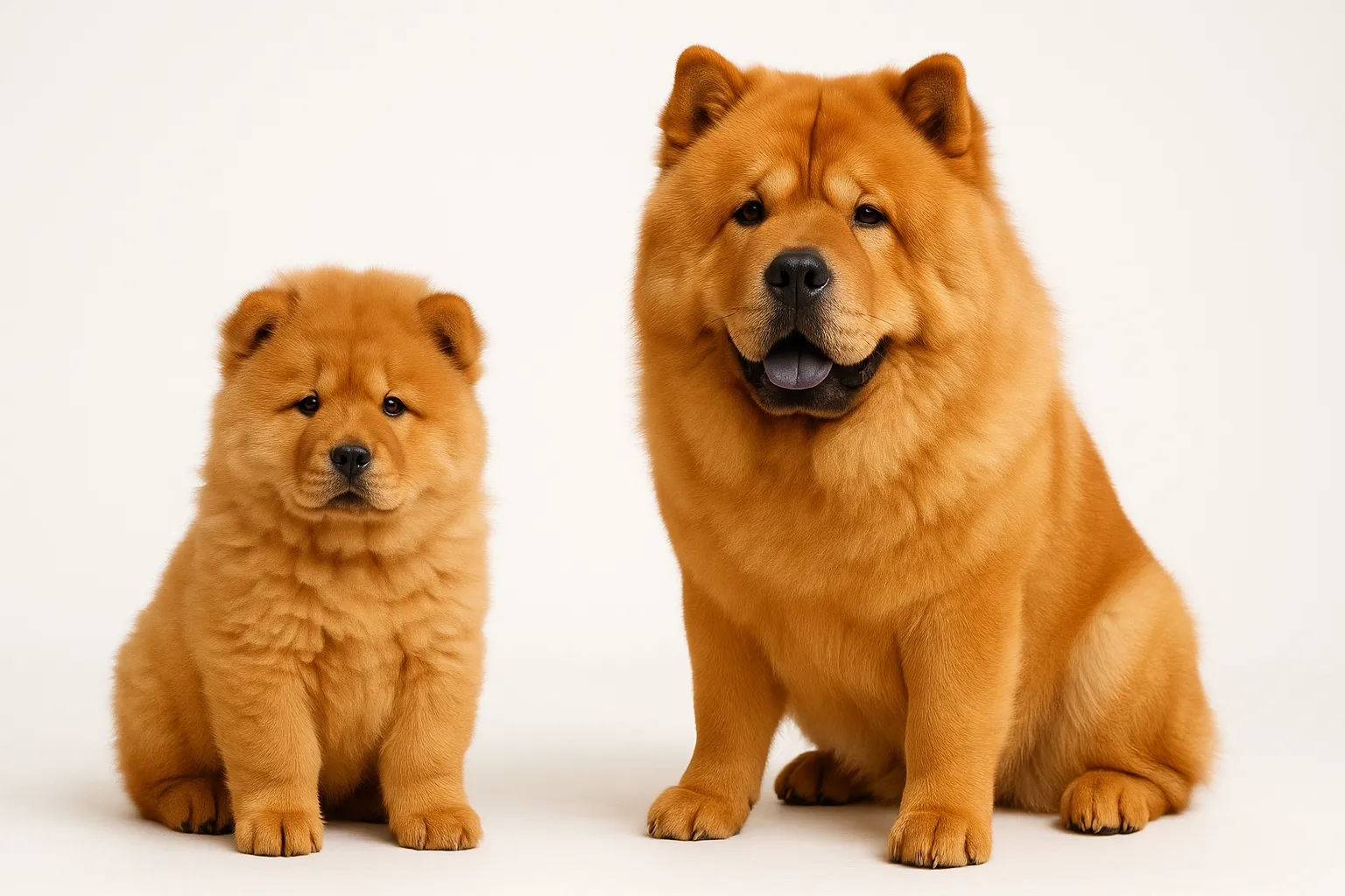
- Chow Chow: significantly elevated risk
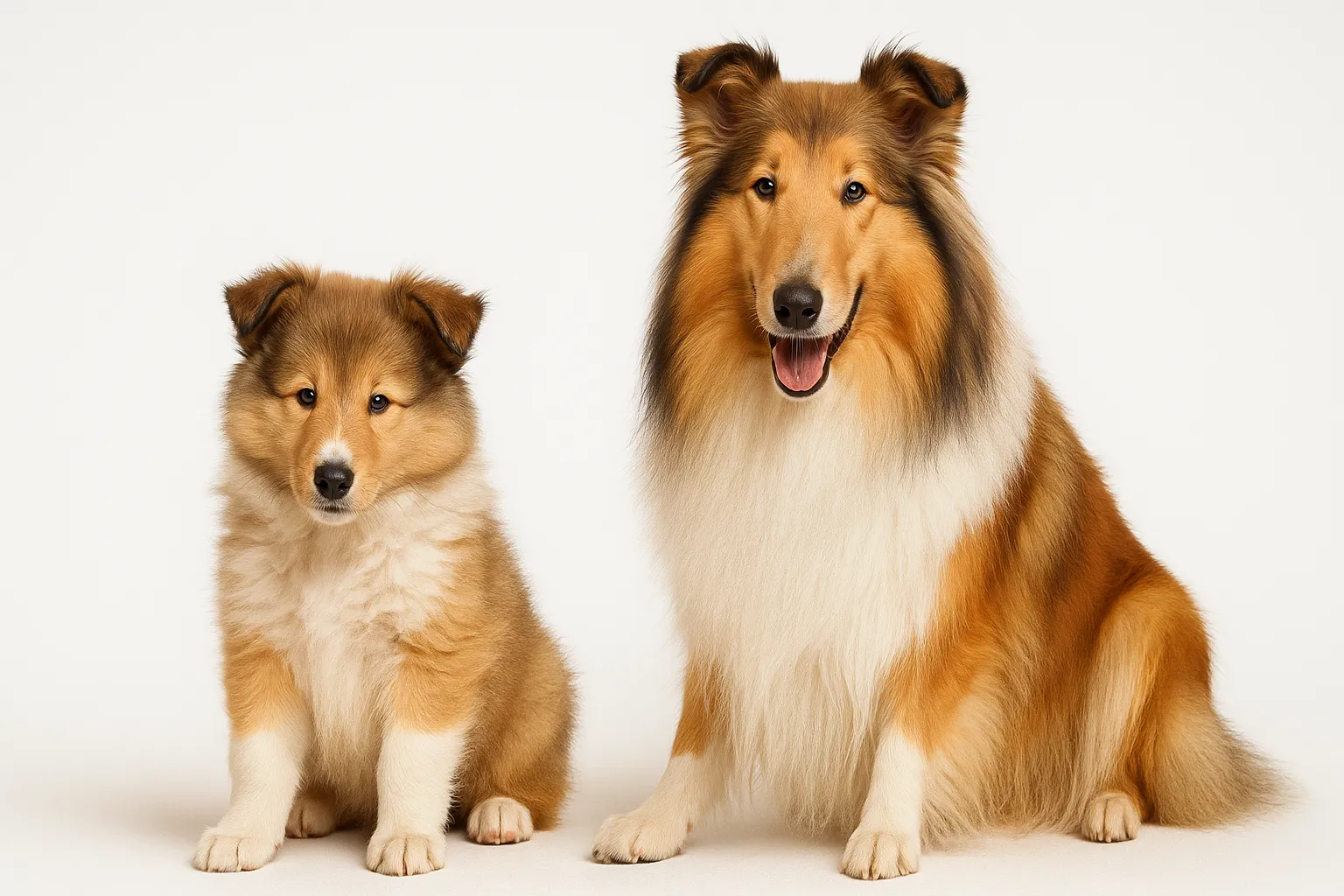
- Collie: documented breed predisposition
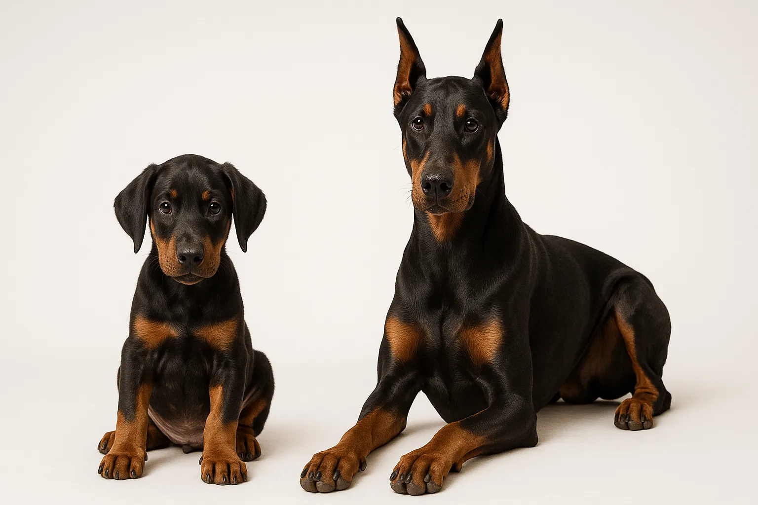
- Doberman Pinscher: increased prevalence
Dachshunds, Finnish Spitz, Newfoundlands, and Schipperkes also appear in the literature. No consistent sex predisposition exists, though some studies report slightly higher incidence in spayed females.
Recognizing Pemphigus
The First Signs — Often Mistaken for Something Else
If your Akita or Chow develops crusty lesions on the nose or cracking footpads that do not resolve with antibiotics, pemphigus deserves investigation.
- Pustules on the face, especially the bridge of the nose and around the eyes
- Crusts and erosions on the ear flaps
- Footpad thickening, fissuring, and crusting — one of the most characteristic early findings
- Depigmentation and crusting of the nose
- Mild itching in some dogs
As It Progresses
- Crusts and erosions spread to the trunk, limbs, and groin
- Thick, adherent crusts that reveal moist, raw tissue when removed
- Footpads develop painful fissures that cause limping
- Secondary bacterial infection with pustular discharge
- Fever, lethargy, and decreased appetite
- Enlarged lymph nodes
Severe, Generalized Disease
- Widespread crusting and erosions covering large body surface areas
- Significant pain and reluctance to move
- Weight loss from chronic protein loss and poor appetite
- Secondary sepsis from bacteria invading through the destroyed skin barrier
What Triggers the Immune Attack
Autoantibodies against desmoglein-1 drive PF. IgG antibodies bind this intercellular adhesion protein, causing superficial skin cells to lose cohesion. Why the immune system begins producing these antibodies remains unknown in most cases.
Genetics set the stage. Strong breed associations point to heritable susceptibility factors, likely involving major histocompatibility complex (MHC) genes that govern immune recognition.
Certain drugs can trigger pemphigus in predisposed dogs. Trimethoprim-sulfadiazine, cephalosporins, and some topical flea/tick preventatives have all been implicated. Drug-induced PF sometimes resolves when the medication is stopped — but not always.
Ultraviolet light may worsen PF in some dogs, particularly lesions on the nose and dorsal muzzle. This association is strongest with pemphigus erythematosus.
Pre-existing skin inflammation may alter immune tolerance and contribute to disease onset in genetically susceptible dogs.
Getting the Diagnosis Right
Skin Biopsy — The Essential Test
Definitive diagnosis requires skin biopsy. The pathologist looks for subcorneal acantholysis (separation of superficial skin cells) with neutrophilic or eosinophilic pustule formation. The diagnostic yield is highest from intact pustules rather than ruptured erosions.
Multiple biopsies from 3-4 sites at different stages of lesion development increase accuracy. Footpad samples, when affected, are particularly diagnostic.
Cytology — A Rapid Preliminary Answer
Pressing a slide against an intact pustule or beneath a freshly removed crust reveals acantholytic keratinocytes — rounded, detached skin cells surrounded by neutrophils. This finding strongly suggests pemphigus and provides a working diagnosis while biopsy results are pending.
Supporting Workup
- Complete blood count: may show elevated neutrophils, eosinophils
- Chemistry panel: baseline before starting immunosuppressive drugs
- Bacterial culture and sensitivity: guides antibiotic selection when secondary infection complicates the picture
- ANA testing: rules out systemic lupus erythematosus
- Drug history review: identifies potential triggers
Prevention — Limited, But Early Recognition Matters
Pemphigus cannot be prevented. The autoimmune process and the triggers that initiate it remain incompletely understood.
What owners can do:
- Avoid known drug triggers in predisposed breeds when alternatives exist
- Minimize unnecessary UV exposure in dogs with facial pemphigus
- Learn the early signs — nasal and footpad crusting in an at-risk breed should prompt investigation, not wait-and-see
- Early diagnosis means lower drug doses needed for control and less tissue damage to heal
Treatment: Suppressing the Immune Attack
Immunosuppressive Drugs
Corticosteroids are the first-line treatment. Prednisone or prednisolone at immunosuppressive doses (2-4 mg/kg/day) is started until clinical remission (typically 2-4 weeks), then tapered gradually over weeks to months to the lowest dose that maintains control.
Azathioprine (2 mg/kg daily, then tapered) is commonly added as a steroid-sparing agent to reduce long-term corticosteroid side effects. Requires CBC monitoring every 2 weeks initially for bone marrow suppression.
Mycophenolate mofetil (10-20 mg/kg twice daily) offers an alternative steroid-sparing approach with fewer side effects than azathioprine in many dogs.
Chlorambucil is reserved for refractory cases, particularly dogs that cannot tolerate azathioprine.
Cyclosporine (5 mg/kg twice daily) may work as adjunctive therapy but is generally less effective as monotherapy for PF.
Topical Treatment
- Tacrolimus (0.1%) applied to localized facial or footpad lesions can reduce the need for systemic drugs
- Gentle wound care: non-adherent dressings, antiseptic soaks for open erosions
- Zinc-free sunscreen for UV-sensitive facial lesions
Fighting Secondary Infection
Bacterial invasion through broken skin is common and can mimic disease flares. Culture-guided systemic antibiotics treat active infection. Routinely culturing new pustules helps distinguish a true PF flare (needs more immunosuppression) from secondary infection (needs antibiotics, not more immune suppression) — a critical distinction that drives completely different treatment decisions.
Nutritional Support
No supplement treats pemphigus directly, but nutritional support complements immunosuppressive management:
- Omega-3 fatty acids at anti-inflammatory doses (50-100 mg/kg/day combined EPA+DHA) may modestly reduce inflammation and support skin barrier repair
- Adequate protein intake supports skin healing and offsets protein lost through weeping lesions
- Vitamin E at moderate doses (400-600 IU daily) provides antioxidant support for skin under inflammatory stress
- Dogs on long-term corticosteroids benefit from calcium-adequate diets to protect bone density
- Probiotics for Dogs may help maintain gut health during immunosuppressive therapy, though evidence specific to pemphigus is lacking
When Your Dog Needs a Vet
Routine monitoring is appropriate for:
- Dogs on stable maintenance immunosuppressive therapy (recheck every 2-4 months)
- Bloodwork monitoring for medication side effects (CBC, chemistry every 2-4 weeks during induction, then every 3-6 months)
Prompt evaluation is needed for:
- New pustules or crusts appearing during maintenance therapy (flare vs. secondary infection — the distinction matters)
- Footpad fissuring causing lameness
- Signs of corticosteroid side effects: excessive thirst/urination, panting, muscle weakness, skin thinning
Emergency evaluation — do not wait:
- Rapidly spreading erosions covering large body surface areas
- Fever with extensive skin lesions (suggesting secondary sepsis)
- Acute illness in an immunosuppressed dog (infections can escalate quickly)
- Severe footpad pain preventing weight-bearing
Related Condition Pathways
Related Breed Longevity Guides
Related Science
- Omega-3 for Dogs: Evidence and Safety
- Supplement Evidence for Dog Longevity
- Evaluating Longevity Supplement Claims for Dogs
Frequently Asked Questions
Is pemphigus foliaceus curable? No. PF is managed, not cured. Some dogs achieve sustained remission on low-dose immunosuppressive therapy, and occasional dogs tolerate complete drug withdrawal. But the majority require lifelong medication. Drug-induced PF may resolve if the triggering medication is identified and stopped.
How long does treatment take to work? Most dogs improve within 2-4 weeks of starting immunosuppressive therapy, with significant remission by 4-8 weeks. Footpad lesions typically heal slowest. Treatment continues well past remission — the drugs taper gradually to the lowest effective dose rather than stopping.
Are pemphigus dogs immunocompromised? Yes. The immunosuppressive therapy necessary to control PF reduces the immune system’s ability to fight infections. Dogs on these drugs should be monitored for signs of illness, kept current on appropriate vaccinations (killed vaccines only — no modified-live vaccines), and evaluated promptly for any new health concern.
Can pemphigus be triggered by vaccines? Case reports have described vaccine-associated pemphigus, though the causal link remains unproven. Some veterinary dermatologists recommend cautious vaccination planning in dogs with pemphigus history: essential vaccines only, no unnecessary boosters. Discuss your dog’s vaccination strategy with a veterinary dermatologist.
Is pemphigus painful? Yes. Active lesions — especially footpad fissures and widespread erosions — cause significant pain. Pain management with appropriate analgesics is part of comprehensive care, particularly during disease flares before immunosuppressive therapy achieves adequate control.
Medical Disclaimer
This content is for educational purposes only and does not constitute veterinary medical advice. Pemphigus requires professional diagnosis through skin biopsy and histopathology. Immunosuppressive therapy carries significant risks and should be managed by a veterinarian, ideally a board-certified veterinary dermatologist, familiar with your dog’s individual case.
References
[1] Olivry T, Chan LS, Xu L, et al. “Novel canine pemphigus foliaceus autoantigens identified by immunoprecipitation.” J Immunol. 2003;170(6):3313-3320. [2] Olivry T. “A review of autoimmune skin diseases in domestic animals: I - Superficial pemphigus.” Vet Dermatol. 2006;17(5):291-305. [3] Mueller RS, Krebs I, Power HT, Fieseler KV. “Pemphigus foliaceus in 91 dogs.” J Am Anim Hosp Assoc. 2006;42(3):189-196. [4] Gomez SM, Morris DO, Rosenbaum MR, Goldschmidt MH. “Outcome and complications associated with treatment of pemphigus foliaceus in dogs: 43 cases (1994-2000).” J Am Vet Med Assoc. 2004;224(8):1312-1316. [5] Bizikova P, Olivry T. “A randomized, double-blinded crossover trial testing the benefit of two antipruritic sprays for the treatment of canine pemphigus foliaceus.” Vet Dermatol. 2016;27(1):50-e16. [6] Scott DW, Miller WH, Griffin CE. Muller and Kirk’s Small Animal Dermatology. 7th ed. Saunders Elsevier; 2013.
Related reads
Related Reading
Continue exploring