Evidence deep dives for Cranial Cruciate Ligament Rupture
Pair mechanism-level evidence with practical protocol context before discussing next steps with your veterinarian.
The $1.3 Billion Knee Problem in Dogs
One day your dog is running across the yard. The next, they are holding up a hind leg and refusing to put weight on it. You might assume they stepped on something, pulled a muscle, or just “tweaked” their knee. But if your dog is a middle-aged Lab, Rottweiler, or Golden, the odds point somewhere more serious: a cranial cruciate ligament rupture.
CCL rupture — the canine equivalent of an ACL tear in humans — is the most common orthopedic condition in dogs and the leading cause of hind-limb lameness in medium to large breeds. American dog owners spend approximately $1.3 billion annually treating it.
Here is the part that catches most owners off guard: unlike human ACL tears, which typically happen during a sudden twist on the sports field, canine CCL rupture is almost always a degenerative process. The ligament weakens over months to years before it finally gives way. That is also why 40-60% of dogs that rupture one knee will rupture the other within 1-2 years. The problem is bilateral. The deterioration is systemic.
Why This Injury Reshapes Your Dog’s Future
When the CCL fails, the stifle (knee) joint loses its primary stabilizer. The tibia slides forward with every step. The meniscus gets damaged. Osteoarthritis begins immediately.
Left untreated, the consequences cascade:
- Severe osteoarthritis develops within 6-12 months
- Mobility declines, activity drops, weight climbs
- The sedentary cycle accelerates other age-related conditions
Surgical stabilization — TPLO or TTA — returns 85-95% of dogs to functional limb use and slows arthritis progression compared to conservative management. But even after successful surgery, the other knee remains at risk. Weight management and controlled exercise stay essential for life.
Which Dogs Are Most Vulnerable
CCL rupture concentrates in certain breeds:
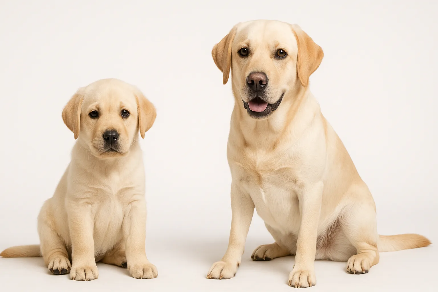
- Labrador Retriever: the single most commonly affected breed, with research suggesting genetic susceptibility to ligament degeneration
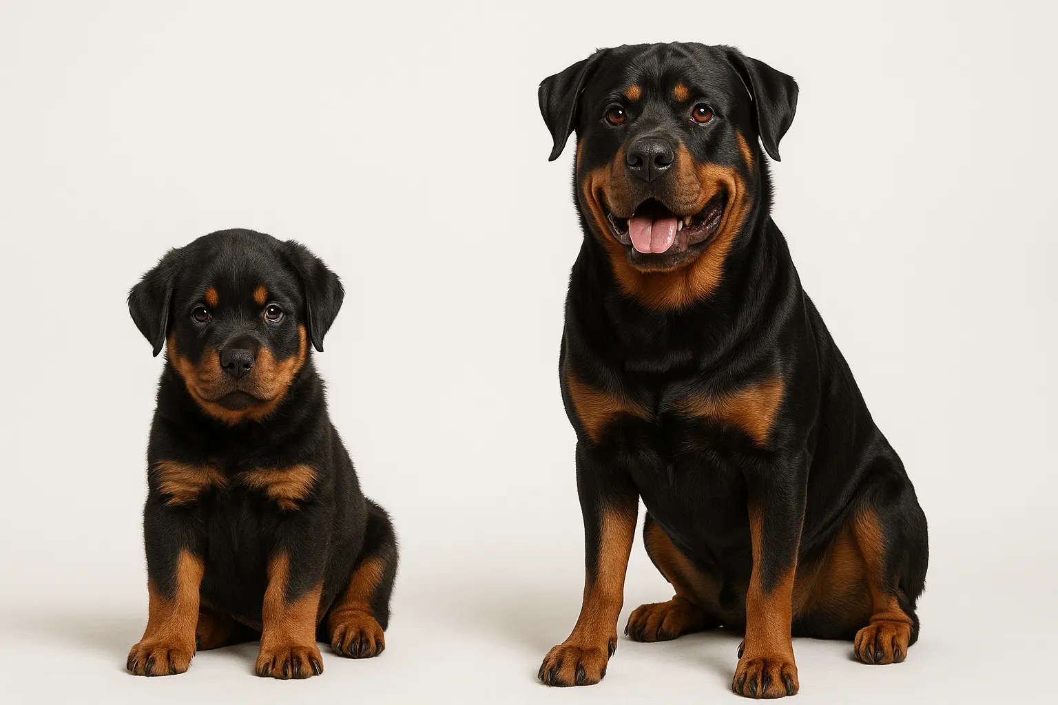
- Rottweiler: significantly over-represented, especially for bilateral disease
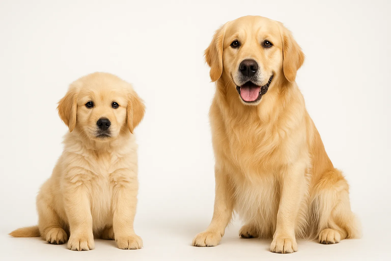
- Golden Retriever: elevated risk consistent with their broader orthopedic disease burden
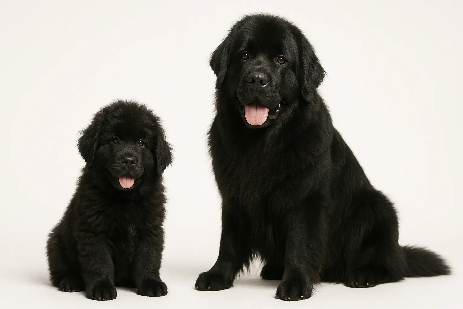
- Newfoundland: high prevalence, often presenting at younger ages
Staffordshire Bull Terriers, West Highland White Terriers, Boxers, and American Pit Bull Terriers also appear frequently. Greyhounds are almost never affected, hinting at protective genetic factors. Dogs weighing over 22 kg (48 lbs) carry substantially higher risk.
One finding that has reshaped veterinary thinking: neutered dogs rupture their CCLs more often than intact dogs. A UC Davis study found that early neutering (before age 1) carried a 2-3x increase in CCL disease risk in several breeds. The mechanism may involve altered body composition, growth plate timing effects on joint conformation, or direct hormonal influences on ligament biology.
Recognizing the Signs
Partial Tear — The Warning Phase
If your dog has started limping after walks but seems fine after resting, a partial CCL tear may already be underway.
- Intermittent hind-limb lameness that worsens with activity
- Stiffness after rest, especially after lying down for a long time
- Reluctance to jump, climb stairs, or fully load the affected leg
- Mild stifle swelling
- Subtle gait shift, favoring the other leg
Complete Rupture
- Sudden, significant hind-limb lameness (often after activity, but not always traumatic)
- Refusal to bear weight on the affected leg
- Sitting with the injured leg kicked out to the side (“sit test” positive)
- Visible knee swelling and thickening
- Thigh muscle atrophy within 2-3 weeks
- Clicking or popping sound during movement (suggests meniscal damage)
Both Knees Affected
- Shifting weight between hind limbs
- Difficulty rising from sitting or lying
- Bunny-hopping gait
- Progressive reduction in activity
- Dogs with bilateral disease are often mistaken for “just getting old” — the lameness is symmetrical, so no single leg stands out
What Causes the Ligament to Fail
Chronic degeneration drives the process. Histopathological studies show that virtually all dogs with CCL rupture have pre-existing degenerative changes in both cruciate ligaments. The ligament does not snap from one bad landing. It erodes from within.
Genetics load the dice. Research has identified candidate genetic loci in Labrador Retrievers and Newfoundlands linked to CCL disease. The heritable component likely involves collagen quality, inflammatory pathways, and joint conformation.
Excess weight accelerates breakdown. Every extra pound increases mechanical load on the stifle and may speed ligament degeneration. The Purina Lifetime Study showed that lean dogs had significantly lower rates of musculoskeletal disease.
Steep tibial plateau angle matters. Dogs with steeper angles experience greater forward thrust forces during weight-bearing, stressing the CCL with every step.
Neutering changes the equation. Multiple studies document increased CCL risk in neutered dogs, particularly those neutered before skeletal maturity. Altered body composition, joint conformation changes, and direct hormonal effects on the ligament all play potential roles.
Pre-existing joint inflammation may accelerate degeneration in some dogs.
How Veterinarians Diagnose CCL Rupture
Hands-On Examination
Cranial drawer test: The vet stabilizes the femur and tries to slide the tibia forward. Any forward movement signals CCL compromise.
Tibial thrust test: Flexing the hock while the stifle is at a standing angle produces forward tibial movement if the CCL has failed. This test works especially well in large-breed dogs where muscle tension can mask the drawer sign.
Palpation: The vet checks for joint swelling, medial buttress (thickened tissue on the inner knee), and thigh muscle atrophy.
Imaging
Stifle radiographs confirm effusion, measure existing arthritis, rule out fractures or bone tumors, and calculate the tibial plateau angle for surgical planning. MRI can identify partial tears and meniscal damage that X-rays miss, though it is not routinely required. Arthroscopy provides direct visualization and is typically performed at the time of surgery.
Reducing the Risk Before It Happens
You cannot eliminate genetic predisposition, but you can modify the factors that accelerate ligament failure.
- Keep your dog lean. Maintaining a body condition score of 4-5/9 throughout life is the single most impactful thing you can do. Excess weight is the biggest modifiable risk factor.
- Control high-impact activity. Repetitive hard fetching on pavement, uncontrolled off-leash sprinting, and sudden directional changes on slippery surfaces create excessive stifle torque. Structured exercise that builds muscle without overloading joints is the better path.
- Build supportive muscle. Strong quadriceps and hamstrings stabilize the stifle dynamically. Exercise protocols by life stage should emphasize balanced muscle development.
- Consider spay/neuter timing carefully. Discuss breed-specific CCL risk data with your veterinarian before deciding on timing.
- Improve traction at home. Carpet runners, area rugs, trimmed nails, and paw wax reduce high-speed sliding on tile, hardwood, and wet grass.
Surgical Options — And When to Choose Each
TPLO: The Gold Standard
Tibial Plateau Leveling Osteotomy is the most commonly performed and best-studied procedure. The surgeon makes a semicircular cut in the proximal tibia and rotates the tibial plateau to neutralize forward thrust. Bone heals over 6-8 weeks.
Outcome: 90-95% of dogs achieve good-to-excellent function. Cost: $3,500-$6,500 per stifle.
TTA: A Comparable Alternative
Tibial Tuberosity Advancement changes the patellar tendon angle to eliminate forward thrust. Published outcomes match TPLO, with some evidence suggesting faster early recovery. Cost: $3,000-$5,500.
Lateral Suture: For Smaller Dogs
An extracapsular suture placed outside the joint mimics CCL function. Less invasive, less expensive ($1,500-$3,000), and well suited for dogs under 15-20 kg. Larger dogs have higher failure rates with this technique.
When Surgery Is Not an Option
Conservative management — strict rest, NSAIDs, physical rehabilitation, weight loss, and joint supplements — may be appropriate for dogs under 15 kg, dogs with high anesthetic risk, or when finances preclude surgery. But in dogs over 15 kg, conservative management rarely produces outcomes comparable to surgery. The stifle stays unstable, and arthritis progresses.
Rehabilitation After Surgery
Structured rehab significantly improves surgical outcomes:
- Weeks 1-2: Strict rest, ice therapy, passive range of motion, short leash walks for bathroom only
- Weeks 3-6: Gradually increasing controlled leash walks, underwater treadmill, therapeutic exercises
- Weeks 7-12: Progressive exercise, strength building, gradual return to normal activity
- 12+ weeks: Radiographic recheck confirming bone healing, then gradual return to full activity
Supporting the Joint Nutritionally
Joint supplements serve as adjuncts — not replacements — for surgical treatment and weight management:
- Omega-3 fatty acids (EPA/DHA from fish oil) at 50-100 mg combined per kg daily: the strongest evidence base among joint supplements for anti-inflammatory benefit
- Glucosamine and chondroitin: moderate evidence for osteoarthritis support; widely used for long-term joint health
- Collagen peptides: emerging evidence for connective tissue support
- MSM: limited evidence but generally safe
- Adequate protein intake to support the muscle mass that stabilizes the joint dynamically
- Weight loss feeding protocol if overweight: achieving lean body condition delivers more joint benefit than any supplement
When Your Dog Needs a Vet
Routine evaluation is appropriate for:
- Annual orthopedic screening in predisposed breeds
- Dogs with a previous CCL rupture (the other knee needs monitoring)
- Mild, intermittent stiffness after exercise in middle-aged large-breed dogs
Prompt evaluation is needed for:
- Persistent hind-limb lameness lasting more than 48 hours
- Progressive lameness worsening over weeks
- Stiffness and difficulty rising in middle-aged large-breed dogs
- Visible stifle swelling
Urgent evaluation is required for:
- Sudden non-weight-bearing lameness in a hind limb
- Acute inability to rise
- Audible popping or clicking from the knee
- Rapidly progressive lameness with significant swelling
Related Condition Pathways
Related Breed Longevity Guides
Related Science and Nutrition
- Canine Obesity and Lifespan Evidence
- Weight Management Protocol for Dogs
- Spay-Neuter Timing and Longevity Context
- Muscle and Mobility Longevity Protocol
- Glucosamine and Chondroitin for Dogs
- Omega-3 Fish Oil for Dogs
Frequently Asked Questions
Does my dog need surgery for a torn CCL? For dogs over 15-20 kg, surgery (TPLO or TTA) delivers significantly better outcomes than conservative management. Without surgery, the stifle remains mechanically unstable, and arthritis progresses steadily. For smaller dogs under 15 kg, conservative management with strict rest and rehabilitation produces acceptable outcomes in many cases.
What is the difference between TPLO and TTA? Both procedures neutralize stifle instability by changing joint biomechanics rather than replacing the ligament. TPLO rotates the tibial plateau; TTA advances the tibial tuberosity. Published outcome studies show similar long-term results. Surgeon experience, individual patient anatomy, and cost typically drive the choice.
Will my dog’s other knee also rupture? Approximately 40-60% of dogs that rupture one CCL will rupture the opposite knee within 1-2 years. Risk rises in overweight dogs and those with steep tibial plateau angles. Weight management and controlled activity are the most effective strategies for protecting the second knee.
How long is recovery after TPLO surgery? Full recovery takes approximately 12-16 weeks. Most dogs bear weight on the surgical leg within 1-2 weeks. Bone healing and return to unrestricted activity occur around 10-12 weeks post-surgery. Structured rehabilitation accelerates recovery and improves the final outcome.
Can weight loss prevent CCL rupture? Weight management is the most impactful modifiable risk factor. It cannot override genetic predisposition, but maintaining lean body condition (BCS 4-5/9) significantly reduces mechanical load on the ligament and may slow degenerative changes. Overweight dogs that achieve a lean condition show measurable reductions in orthopedic disease progression.
Medical Disclaimer
This content is for educational purposes only and does not constitute veterinary medical advice. Cranial cruciate ligament disease requires professional diagnosis and treatment planning by a veterinarian, ideally in consultation with a board-certified veterinary surgeon (ACVS). Surgical decisions, rehabilitation protocols, and pain management should be individualized for your dog.
References
[1] Wilke VL, Robinson DA, Evans RB, et al. “Estimate of the annual economic impact of treatment of cranial cruciate ligament injury in dogs in the United States.” J Am Vet Med Assoc. 2005;227(10):1604-1607. [2] Witsberger TH, Villamil JA, Schultz LG, et al. “Prevalence of and risk factors for hip dysplasia and cranial cruciate ligament deficiency in dogs.” J Am Vet Med Assoc. 2008;232(12):1818-1824. [3] Slocum B, Slocum TD. “Tibial plateau leveling osteotomy for repair of cranial cruciate ligament rupture in the canine.” Vet Clin North Am Small Anim Pract. 1993;23(6):1169-1182. [4] Torres de la Riva G, Hart BL, Farver TB, et al. “Neutering dogs: effects on joint disorders and cancers in Golden Retrievers.” PLoS One. 2013;8(2):e55937. [5] Comerford EJ, Smith K, Hayashi K. “Update on the aetiopathogenesis of canine cranial cruciate ligament disease.” Vet Comp Orthop Traumatol. 2011;24(2):91-98. [6] Christopher SA, Beetem J, Cook JL. “Comparison of long-term outcomes associated with three surgical techniques for treatment of cranial cruciate ligament disease in dogs.” Vet Surg. 2013;42(3):329-334.
Related reads
Related Reading
Continue exploring