Evidence deep dives for Cranial Cruciate Ligament Tear (CCL/ACL)
Pair mechanism-level evidence with practical protocol context before discussing next steps with your veterinarian.
The Most Common Orthopedic Surgery in Dogs
Your dog was running in the yard and suddenly pulled up lame on a hind leg. They are holding the leg up or barely touching the toe down. Within an hour, the knee is swollen. Within a day, they are limping noticeably but still using the leg. Within a week, they seem better. Within a month, the lameness returns and is worse than before.
This pattern is the signature of a cranial cruciate ligament (CCL) tear, the canine equivalent of the ACL tear in human athletes. It is the single most common orthopedic condition requiring surgery in dogs, accounting for an estimated $1.3 billion in annual veterinary spending in the United States alone.
But there is a critical difference between canine CCL disease and human ACL tears. In humans, ACL rupture is typically an acute sports injury caused by a specific traumatic event. In dogs, CCL rupture is almost always the end result of a chronic degenerative process. The ligament weakens progressively over months or years before it finally fails, sometimes during normal activity. This distinction matters because it means the other knee’s ligament is likely undergoing the same degenerative process, and 40-60% of dogs that rupture one CCL will rupture the opposite side within 1-2 years.
Anatomy and Why It Fails
The stifle (knee) joint is stabilized by two cruciate ligaments that cross inside the joint:
- The cranial cruciate ligament (CCL) prevents the tibia from sliding forward relative to the femur and limits internal rotation and hyperextension
- The caudal cruciate ligament prevents backward tibial displacement and is rarely injured
The CCL bears tremendous mechanical load during normal activity. When a dog pushes off the ground, the tibial plateau (the top surface of the shin bone) slopes backward. Without an intact CCL, the femur slides backward on the sloped tibial surface, and the tibia thrusts forward. This abnormal motion is called cranial tibial thrust and is the fundamental mechanical problem that CCL surgery aims to correct.
Why Degeneration, Not Trauma, Is the Typical Cause
Research has demonstrated that most canine CCL tears result from progressive ligament degeneration rather than acute injury. Histological studies of partially torn CCLs show:
- Loss of normal collagen fiber organization
- Decreased cellularity and altered cell morphology
- Chondroid metaplasia (cartilage-like changes in ligament tissue)
- Inflammatory infiltrates
These degenerative changes weaken the ligament over time until it can no longer withstand normal mechanical loads. The rupture may occur during vigorous activity, but the ligament was already compromised. This is why CCL tears commonly occur in middle-aged dogs during routine activity rather than during dramatic athletic events.
Multiple factors drive this degeneration:
- Genetics: Breed-specific differences in stifle conformation and ligament biology
- Tibial plateau angle: Steeper tibial plateau angles increase the mechanical load on the CCL
- Body weight: Excess weight accelerates ligament degeneration. See obesity
- Immune-mediated factors: Lymphocytic-plasmacytic inflammation has been documented in degenerating CCLs, suggesting an immune component
- Neutering: Studies show that early neutering (before sexual maturity) increases CCL rupture risk, potentially through effects on musculoskeletal development
Which Breeds Are Most Affected
CCL disease has a strong breed predisposition:
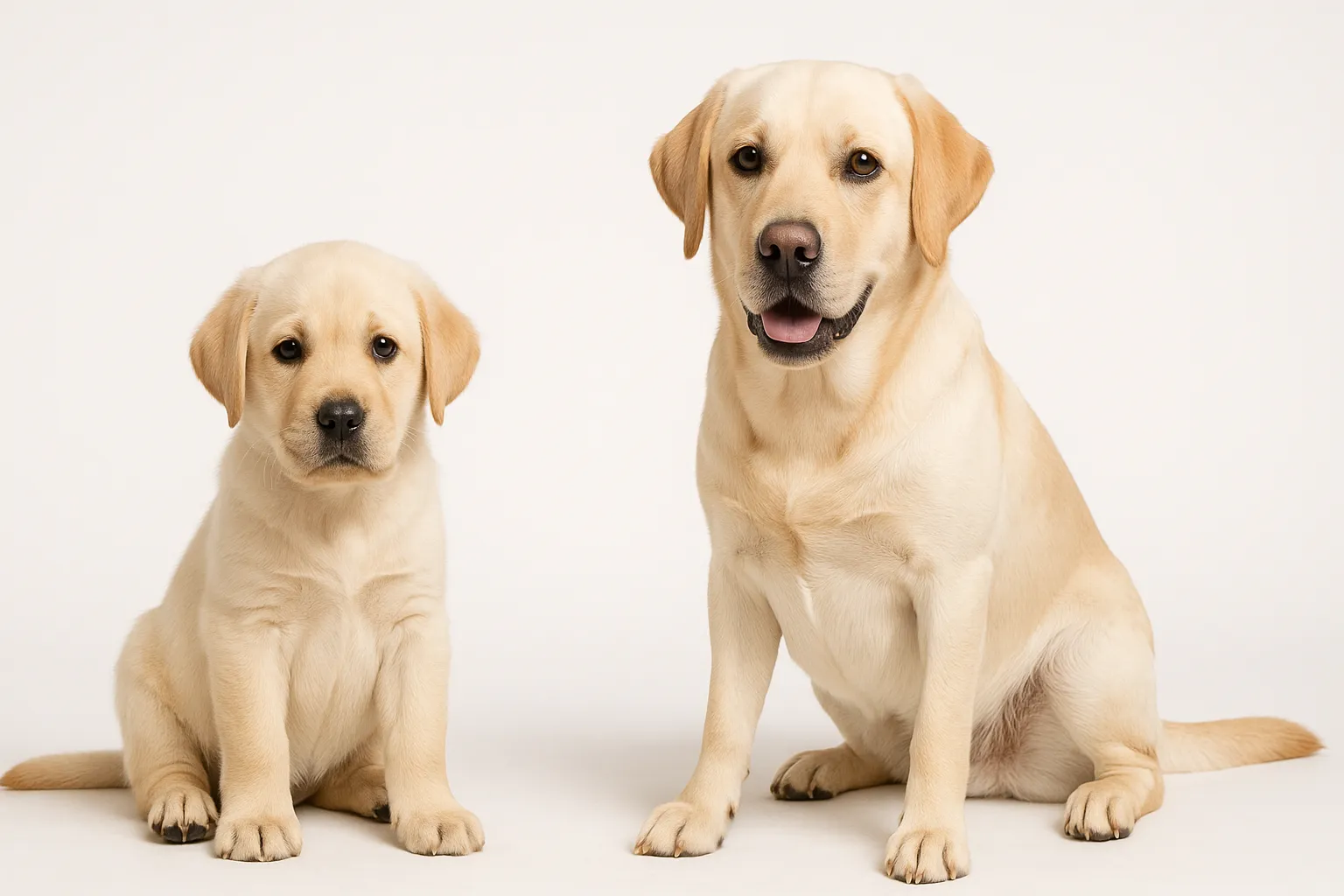
- Labrador Retriever — the breed most commonly requiring CCL surgery, driven by genetics, activity level, and obesity predisposition
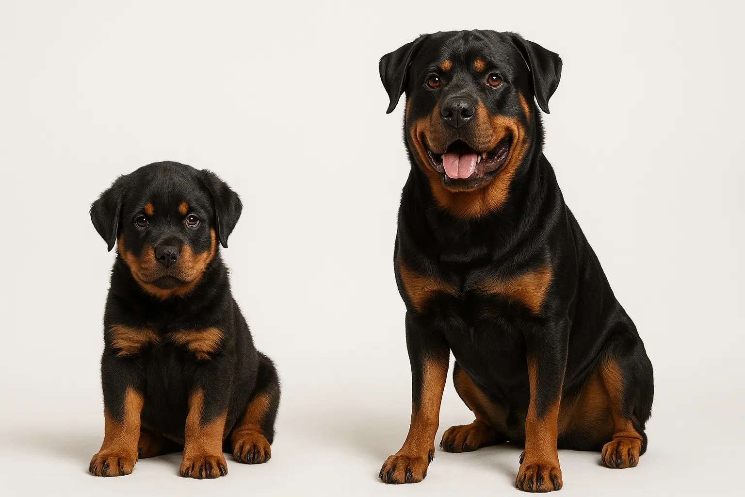
- Rottweiler — steep tibial plateau angle, heavy body weight
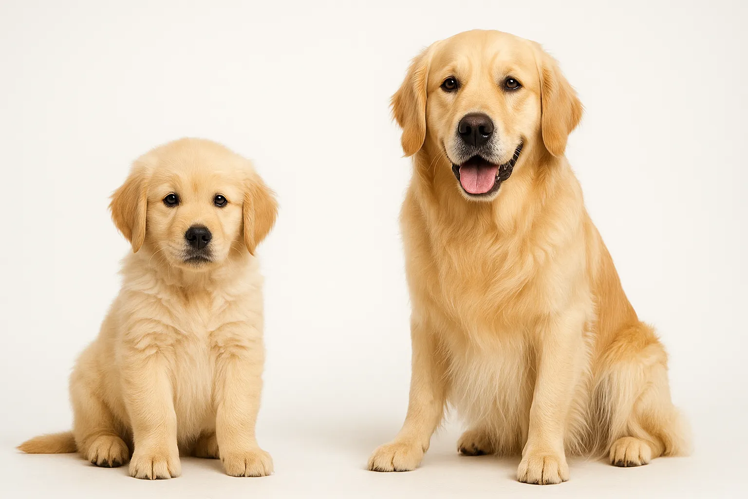
- Golden Retriever — genetic predisposition, active lifestyle
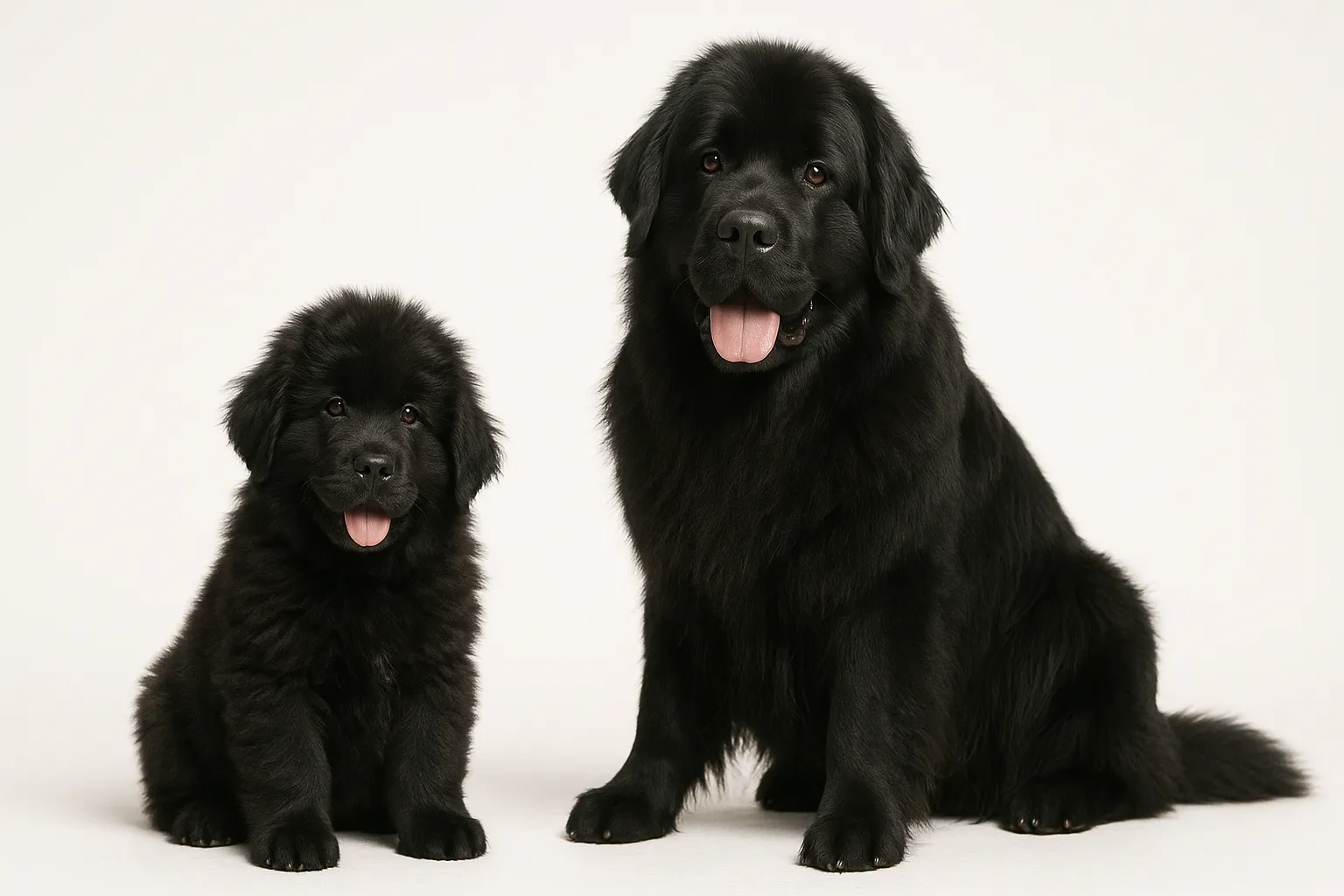
- Newfoundland — heavy body weight, steep tibial plateau angle. Studies show a lifetime CCL rupture incidence as high as 22% in Newfoundlands
- Saint Bernard — giant breed with mechanical predisposition
- Staffordshire Bull Terrier — documented breed predisposition
- West Highland White Terrier — despite small size, has a recognized predisposition
While large and giant breeds are most commonly affected, CCL disease occurs across the size spectrum. Small breed dogs can also rupture their CCL but may respond better to conservative management than larger dogs.
Signs and Symptoms
Acute Partial Tear
- Sudden onset of hind leg lameness, often during or after activity
- The dog may still bear weight but with a noticeable limp
- Stifle joint effusion (swelling)
- Pain on stifle manipulation, particularly when the joint is fully extended
- Mild instability detectable on veterinary examination
- Lameness that improves with rest but returns with activity
Complete Tear
- Sudden, severe lameness (often non-weight-bearing initially)
- Marked stifle joint swelling
- Positive cranial drawer sign (the tibia can be manually displaced forward relative to the femur)
- Positive tibial thrust test
- Sitting with the affected leg extended to the side (“lazy sit”)
- Difficulty rising from a lying position
- Muscle atrophy of the affected thigh (develops over weeks)
Chronic CCL Disease
Over time, an unstable stifle develops progressive osteoarthritis:
- Chronic, persistent lameness that worsens with activity and cold weather
- Joint thickening from fibrosis and osteophyte formation
- Progressive muscle atrophy
- Decreased range of motion
- Pain with joint manipulation
- Meniscal damage (clicking or locking sensation during movement)
The Contralateral Knee
In dogs with unilateral CCL rupture, the opposite knee bears disproportionate load and its CCL is often already degenerating. Approximately 40-60% of dogs will rupture the second CCL within 1-2 years. Monitoring the contralateral stifle is an essential part of long-term management.
Diagnosis
Physical Examination
- Cranial drawer test: With the dog sedated, the examiner stabilizes the femur and attempts to slide the tibia forward. Forward displacement indicates CCL disruption. This test may be negative in partial tears or in dogs with significant muscle tension (hence the need for sedation)
- Tibial thrust test: With the stifle at standing angle, the hock is flexed, generating a cranial thrust of the tibia. A positive test confirms CCL insufficiency
- Stifle effusion: Palpable fluid accumulation in the joint
- Medial buttress: A firm swelling on the inside of the stifle, representing periarticular fibrosis from chronic instability
Imaging
- Radiographs: Do not show the ligament itself but reveal joint effusion, osteophyte formation (arthritis), and changes consistent with chronic instability. Pre-surgical radiographs measure the tibial plateau angle, which guides surgical planning
- MRI: Can visualize the ligament directly and identify partial tears, meniscal damage, and early cartilage changes. Not always necessary but useful in equivocal cases
- Ultrasound: Occasionally used to assess the integrity of the ligament and menisci
Meniscal Evaluation
The menisci (C-shaped cartilage pads between the femur and tibia) serve as shock absorbers. Approximately 40-60% of dogs with CCL rupture have concurrent meniscal damage (most commonly the medial meniscus). Meniscal injury causes pain, clicking, and can impair surgical outcomes if not addressed. Meniscal evaluation occurs during surgery.
Treatment Options
Surgical Treatment (Recommended for Most Dogs Over 15 kg)
Surgery is the standard of care for CCL rupture in medium, large, and giant breed dogs. Multiple techniques exist, but all aim to restore stifle stability:
TPLO (Tibial Plateau Leveling Osteotomy): The current gold standard. A semicircular cut is made in the top of the tibia, and the tibial plateau is rotated to reduce its angle, eliminating the tibial thrust that occurs during weight-bearing. The bone is stabilized with a plate and screws. TPLO produces consistent, excellent functional outcomes in multiple studies, with 90-95% of dogs returning to normal or near-normal function.
TTA (Tibial Tuberosity Advancement): The tibial tuberosity (the front of the shin bone where the patellar tendon attaches) is advanced forward, changing the angle of the patellar tendon force vector to neutralize cranial tibial thrust. Results are comparable to TPLO in most studies.
Lateral Suture Stabilization (Extracapsular Repair): A strong synthetic suture is placed outside the joint to replace the CCL’s function. Simpler and less expensive than TPLO/TTA. Best results are in dogs under 15 kg. In larger dogs, the suture may stretch or fail over time, though periarticular fibrosis provides secondary stability.
Tight Rope Technique: A strong braided suture passed through bone tunnels provides extracapsular stabilization. Similar concept to lateral suture but with a different fixation approach.
Which surgery is best? For dogs over 15 kg, TPLO and TTA produce the most consistent long-term outcomes. For dogs under 15 kg, lateral suture or Tight Rope techniques are reasonable alternatives. The surgeon’s experience with a given technique is often more important than the specific technique chosen.
Conservative Management
Non-surgical management may be appropriate for:
- Small dogs under 15 kg (which can often stabilize with fibrosis)
- Dogs with significant anesthetic risk
- Financial constraints (CCL surgery typically costs $2,500-$6,000 per knee)
- Partial tears in any size dog (some partial tears stabilize with rest and rehabilitation)
Conservative management includes:
- Strict rest: 6-8 weeks of leash-only exercise, no running, jumping, or off-leash activity
- Weight management: Essential. Even modest weight reduction decreases stifle load significantly
- Physical rehabilitation: Controlled exercises, hydrotherapy, therapeutic ultrasound, and PROM (passive range of motion) exercises maintain muscle mass and joint mobility
- NSAIDs: Carprofen, meloxicam, or deracoxib for pain and inflammation control
- Stifle brace: Custom orthotic braces can provide external stability during healing
- Joint supplements: Omega-3 fatty acids, glucosamine, chondroitin (see below)
Many small dogs managed conservatively develop functional stability through periarticular fibrosis within 2-3 months, though some degree of osteoarthritis progression is expected. Large dogs managed conservatively typically fare less well, with progressive lameness and arthritis.
Post-Surgical Rehabilitation
Rehabilitation after CCL surgery is as important as the surgery itself:
- Weeks 1-2: Strict rest, ice therapy, passive range of motion exercises, short controlled leash walks (5 minutes, 3-4 times daily)
- Weeks 3-8: Gradual increase in leash walk duration, underwater treadmill or swimming, therapeutic exercises for muscle rebuilding
- Weeks 8-16: Progressive return to activity with continued rehabilitation. Most dogs are walking well by 8 weeks and approaching full function by 12-16 weeks
- Bone healing: TPLO bone plate removal is usually not necessary unless infection or plate irritation occurs
Supplement Evidence
Joint supplements are commonly used both before and after CCL surgery:
- Omega-3 fatty acids (fish oil): The strongest evidence among supplements. Anti-inflammatory EPA and DHA at 50-100 mg combined per kg body weight daily reduce joint inflammation and may slow osteoarthritis progression. See Omega-3 Fatty Acids for Dogs
- Glucosamine and chondroitin: Widely used for osteoarthritis. Evidence quality is moderate; some studies show improved comfort and function, others show no significant benefit over placebo. See Glucosamine and Chondroitin for Dogs
- Polysulfated glycosaminoglycan (Adequan): Injectable product with some evidence for modifying osteoarthritis progression
- Green-lipped mussel extract: Contains omega-3 fatty acids and glycosaminoglycans. Limited but promising evidence
Supplements are adjunctive. They support but do not replace weight management, appropriate exercise, pain medication, and surgical stabilization when indicated.
Prevention Considerations
True prevention of CCL disease is difficult because the degenerative process has a significant genetic component. However, modifiable factors include:
- Weight management: The single most impactful modifiable factor. Lean dogs put less mechanical load on the CCL and develop less severe osteoarthritis if rupture occurs
- Appropriate exercise: Build and maintain strong hind limb musculature through regular, consistent exercise. Avoid the “weekend warrior” pattern of inactivity followed by intense bursts
- Avoid excessive impact: Repetitive high-impact activities (dock diving, agility at high intensity, disc catching) increase mechanical load on the CCL
- Consider spay/neuter timing: Discuss the potential musculoskeletal implications of early sterilization with your veterinarian, particularly in large breed dogs
- Monitor the other knee: If one CCL has been treated, proactive monitoring and weight management for the contralateral knee is essential
Related Condition Pathways
Related Breed Longevity Guides
When to Seek Veterinary Care
Routine evaluation is appropriate for:
- Mild, intermittent hind leg lameness that resolves with rest
- Monitoring the contralateral knee in a dog with previous CCL surgery
- Scheduled post-surgical follow-up
Urgent evaluation is needed for:
- Sudden severe hind leg lameness (non-weight-bearing)
- Rapid onset of stifle swelling
- Any hind leg lameness that persists for more than 48 hours
- Worsening lameness in a knee that was previously treated surgically (possible meniscal tear or implant complication)
- Bilateral hind leg lameness (both knees affected simultaneously)
Early diagnosis of partial tears allows for intervention before the ligament completely fails, potentially offering more treatment options and better outcomes.
Frequently Asked Questions
Does my dog definitely need surgery? Not necessarily. Small dogs under 15 kg often stabilize with conservative management (strict rest, weight management, rehabilitation) through periarticular fibrosis. For dogs over 15 kg, surgery produces consistently better functional outcomes than conservative management. The decision involves weighing the dog’s size, activity level, response to rest, and individual circumstances.
Which surgery is best for my dog? TPLO and TTA produce the most reliable long-term results for medium to large dogs. For small dogs, lateral suture or Tight Rope techniques are effective alternatives. The surgeon’s experience and comfort with a specific technique often matters more than the technique itself. Ask your surgeon which procedure they perform most frequently and their complication rates.
Will my dog’s other knee tear too? The risk of contralateral CCL rupture is approximately 40-60% within 1-2 years of the first tear. This is because the same degenerative process is likely occurring in both ligaments. Weight management and maintaining muscle mass through regular controlled exercise may reduce this risk.
How long is recovery from CCL surgery? Most dogs are walking comfortably on the operated leg within 4-8 weeks of surgery. Full recovery, including return to athletic activities, takes 4-6 months. The bone (in TPLO) heals within 8-12 weeks, and rehabilitation continues throughout the recovery period. Rushing the recovery increases complication risk.
Can a torn CCL heal on its own? A completely torn CCL does not heal or reattach in dogs. The joint may stabilize through scar tissue formation (periarticular fibrosis), particularly in small dogs, but the ligament itself does not regenerate. Partial tears may or may not progress to complete tears; some stabilize with strict rest, while others eventually complete.
Is CCL surgery worth the cost? For medium to large dogs, CCL surgery typically produces substantial improvement in comfort, function, and quality of life. Untreated CCL rupture leads to progressive osteoarthritis, chronic pain, muscle atrophy, and compensatory injury to the other knee. Studies show significantly better functional outcomes in surgically treated dogs compared to conservatively managed dogs of similar size.
At what age does CCL disease typically occur? Most CCL tears occur between ages 4-8, though they can occur at any age. Young dogs (under 2 years) with CCL tears often have a strong genetic or conformational predisposition. Senior dogs may tear ligaments that have undergone extensive age-related degeneration. The middle-aged peak reflects the intersection of accumulated degeneration with still-active lifestyles.
Medical Disclaimer
This guide is informational and does not replace in-person veterinary diagnosis or treatment. CCL injury requires professional evaluation to determine the extent of ligament damage, assess meniscal integrity, and discuss the most appropriate treatment approach for your individual dog. If your dog develops sudden, severe hind leg lameness, seek veterinary evaluation promptly.
References
[1] Wilke VL, et al. “Estimate of the annual economic impact of treatment of cranial cruciate ligament injury in dogs in the United States.” J Am Vet Med Assoc. 2005;227(10):1604-1607. [2] Hayashi K, et al. “Histologic changes in ruptured canine cranial cruciate ligament.” Vet Surg. 2003;32(3):269-277. [3] Bergh MS, et al. “Systematic review of surgical treatments for cranial cruciate ligament disease in dogs.” J Am Anim Hosp Assoc. 2014;50(5):315-321. [4] Slocum B, Slocum TD. “Tibial plateau leveling osteotomy for repair of cranial cruciate ligament rupture in the canine.” Vet Clin North Am Small Anim Pract. 1993;23(6):1169-1187. [5] Muir P, et al. “Contralateral cruciate survival in dogs with unilateral non-contact cranial cruciate ligament rupture.” PLoS One. 2011;6(10):e25331. [6] Torres de la Riva G, et al. “Neutering dogs: effects on joint disorders and cancers in golden retrievers.” PLoS One. 2013;8(2):e55937. [7] Wucherer KL, et al. “Short-term and long-term outcomes for overweight dogs with cranial cruciate ligament rupture treated surgically or nonsurgically.” J Am Vet Med Assoc. 2013;242(10):1364-1372.
Related reads
Related Reading
Continue exploring