Evidence deep dives for Myasthenia Gravis
Pair mechanism-level evidence with practical protocol context before discussing next steps with your veterinarian.
The Disease Where Muscles Fail a Little More With Every Step
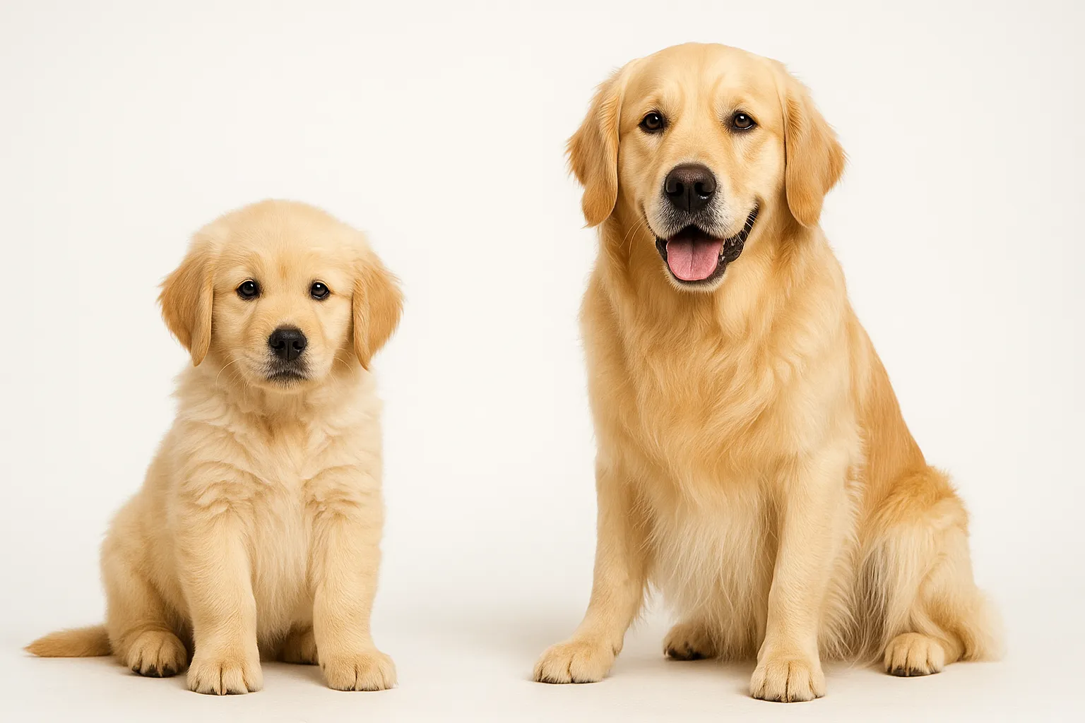
Your Golden Retriever starts a walk looking perfectly normal. Ten minutes later, the legs buckle. After a few minutes of rest, the dog gets up and walks again — until the legs give out once more. This exercise-triggered, rest-responsive weakness is the hallmark of myasthenia gravis (MG), and it is unlike any other cause of weakness in dogs.
MG is an autoimmune disorder in which antibodies attack acetylcholine receptors at the neuromuscular junction — the critical handoff point where nerve signals tell muscles to contract. With fewer functional receptors, signals degrade with repeated use. The muscles work initially, then progressively fail.
About 90% of canine MG cases are acquired (immune-mediated), developing in adult dogs. The remaining 10% are congenital, caused by inherited receptor deficiency that shows up in puppyhood. Acquired MG follows a striking bimodal age pattern: the first peak hits between ages 1-4, the second after age 9. This two-peak distribution likely reflects different immunological triggers at different life stages.
MG presents in three clinical forms:
- Focal MG: weakness confined to specific muscle groups — most commonly the esophageal muscles (causing megaesophagus), throat muscles (causing swallowing difficulty), or facial muscles
- Generalized MG: exercise-induced limb and trunk weakness, often with concurrent megaesophagus
- Acute fulminating MG: rapid-onset, severe weakness affecting respiratory muscles — a medical emergency
Why This Disease Demands Respect
The most dangerous complication is not the muscle weakness itself. It is what happens when the esophageal muscles fail.
Megaesophagus — a dilated, nonfunctional esophagus — develops in roughly 80% of dogs with generalized MG and a significant proportion of those with focal MG. When the esophagus cannot move food into the stomach, food and liquid pool and regurgitate. And regurgitated material can be inhaled into the lungs, causing aspiration pneumonia. That complication is the leading cause of death in dogs with MG.
The prognosis has improved substantially with modern treatment. Spontaneous remission occurs in an estimated 47-88% of dogs with acquired MG, typically within 6-18 months. But the risk of fatal aspiration pneumonia during the active disease period makes early diagnosis and rigorous feeding management the difference between survival and loss.
Breeds at Elevated Risk
Acquired MG concentrates in certain breeds:
- Golden Retriever: one of the most commonly affected, consistent with the breed’s broader predisposition to immune-mediated diseases
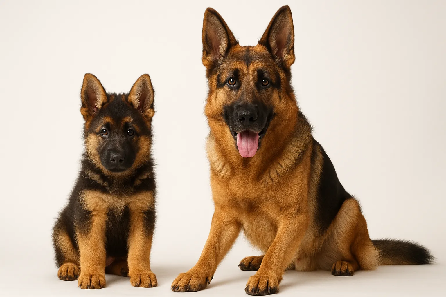
- German Shepherd: over-represented in MG case series
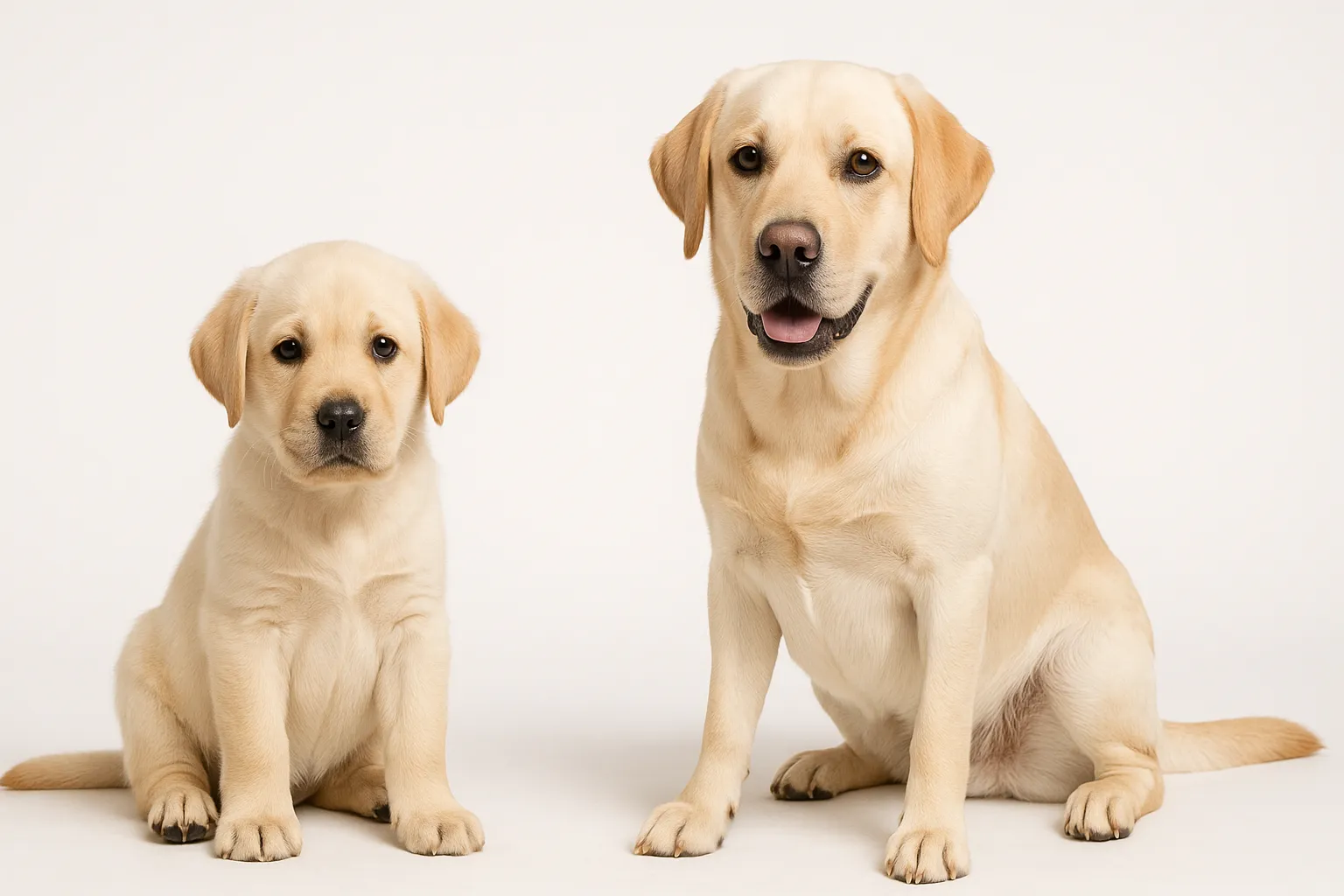
- Labrador Retriever: significant breed predisposition
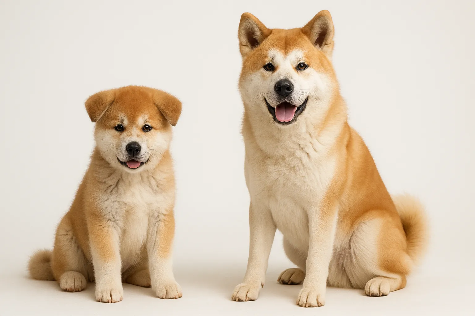
- Akita: elevated risk, particularly for autoimmune conditions
Congenital MG has been documented in Jack Russell Terriers, English Springer Spaniels, Smooth Fox Terriers, and several other breeds. No sex predisposition exists for acquired MG.
What Owners See
Focal MG — It May Look Like a Swallowing Problem
If your dog has started regurgitating undigested food, seemingly without nausea or retching, MG-related megaesophagus may be the cause.
- Regurgitation (passive, tubular expulsion of undigested food — distinct from vomiting)
- Difficulty swallowing (dysphagia)
- Excessive drooling
- Nasal discharge from food entering the nasal passages during swallowing attempts
- Changes in bark quality or a weakened voice
Generalized MG — The Walking-Then-Collapsing Pattern
- Exercise intolerance that worsens with activity and improves after rest
- Progressive limb weakness during walks, sometimes collapsing after moderate exertion
- Short, choppy gait
- Difficulty holding the head up (ventroflexion)
- Regurgitation from concurrent megaesophagus
- Drooping eyelids (palpebral weakness)
Acute Fulminating MG — A True Emergency
- Rapid-onset severe weakness
- Inability to stand or walk
- Respiratory distress from respiratory muscle failure
- Inability to swallow
- This form requires immediate veterinary intervention
What Causes MG
The autoimmune mechanism: In acquired MG, the immune system produces antibodies against nicotinic acetylcholine receptors (AChR). These antibodies degrade the receptors, trigger complement-mediated damage, and physically block acetylcholine from binding. The result: neuromuscular transmission fails proportionally to how many receptors have been lost.
Thymic tumors: Thymoma (a tumor of the thymus gland) is found in approximately 3-5% of dogs with acquired MG. The thymus helps regulate immune tolerance, and thymic abnormalities can trigger or sustain the autoimmune attack on AChR. Every dog diagnosed with MG should have chest imaging to check for thymoma.
Congenital receptor deficiency: Congenital MG stems from genetic mutations affecting AChR expression or function. Most documented breeds show autosomal recessive inheritance. Unlike acquired MG, the congenital form is not immune-mediated.
Broader immune dysfunction: Dogs with MG show increased rates of concurrent autoimmune conditions — hypothyroidism, immune-mediated hemolytic anemia — suggesting the immune dysregulation extends beyond the neuromuscular junction.
Confirming the Diagnosis
The Definitive Test: AChR Antibody Titer
Serum acetylcholine receptor antibody measurement is the gold standard for diagnosing acquired MG. Sensitivity runs approximately 85-90% for generalized MG, with very high specificity. The test is performed by the Comparative Neuromuscular Laboratory at UC San Diego. Results take 5-7 business days.
The Quick Clinical Test: Edrophonium Challenge
Intravenous edrophonium chloride (a short-acting anticholinesterase) produces rapid, dramatic — but temporary — improvement in muscle strength if MG is present. This test provides immediate clinical information but does not replace antibody testing. It requires veterinary supervision with atropine on hand for adverse reactions.
Chest Imaging
- Thoracic radiographs: check for megaesophagus (dilated esophagus filled with gas or food), aspiration pneumonia, and anterior mediastinal mass (thymoma)
- CT scan: indicated if radiographs suggest thymoma
Supporting Tests
- Complete blood count and chemistry panel (baseline health)
- Thyroid panel (screens for concurrent autoimmune thyroiditis)
- Electromyography (EMG): decremental response on repetitive nerve stimulation is characteristic of neuromuscular junction disease
Prevention — Limited but Possible in Specific Areas
MG itself cannot be prevented. The autoimmune and congenital forms are not linked to modifiable risk factors.
What can be prevented are the complications:
- Owners of predisposed breeds should recognize the key signs — unexplained regurgitation, exercise intolerance — and seek prompt evaluation
- Every dog diagnosed with megaesophagus should be tested for MG, because MG is one of the few treatable underlying causes
- Proper feeding management in dogs with megaesophagus prevents the life-threatening complication of aspiration pneumonia
Treatment: Buying Time for Remission
Anticholinesterase Therapy
Pyridostigmine bromide (Mestinon) is the first-line medication. It inhibits the enzyme that breaks down acetylcholine at the neuromuscular junction, increasing the amount of acetylcholine available for the remaining functional receptors. Starting dose: 0.5-3 mg/kg orally every 8-12 hours, titrated to clinical response.
Side effects include GI symptoms (diarrhea, drooling, vomiting) from cholinergic stimulation. Dose adjustments should be gradual.
Immunosuppressive Therapy
For dogs that do not respond fully to pyridostigmine, immunosuppressive drugs target the root autoimmune process:
- Mycophenolate mofetil: increasingly preferred as the first-line immunosuppressive due to fewer side effects; 10-20 mg/kg twice daily
- Azathioprine: 2 mg/kg daily initially, then tapered; requires CBC monitoring for bone marrow suppression
- Corticosteroids: used cautiously because prednisone can initially worsen muscle weakness for 3-5 days; if used, treatment starts at low doses (0.5 mg/kg) with gradual escalation
Megaesophagus Management — The Most Important Part
This is where owners save their dog’s life, one meal at a time:
- Feed from an elevated position (Bailey chair or elevated feeding station) so gravity assists esophageal transit
- Keep your dog upright for 15-30 minutes after every meal
- Feed small, frequent meals in the consistency your individual dog handles best — some do better with gruel, others with formed meatballs
- Watch continuously for signs of aspiration pneumonia: cough, nasal discharge, fever, lethargy
Thymoma Removal
If imaging reveals a thymoma, surgical removal (thymectomy) may improve or resolve MG in some dogs.
Nutrition and Feeding Challenges
No supplement treats or improves myasthenia gravis. Nutritional management revolves around the unique feeding challenges created by megaesophagus:
- Calorie-dense foods reduce the volume needed per meal, minimizing esophageal burden
- Experiment with liquid or gruel-consistency diets if solid food triggers regurgitation
- Bone broth provides calorie-dense liquid nutrition that may be easier for dogs with megaesophagus to swallow safely
- Ensure adequate hydration — dogs with megaesophagus often struggle to drink normally
- Adapt feeding guide principles to elevated positioning and increased meal frequency
- Omega-3 fatty acids provide systemic anti-inflammatory support that may complement immunosuppressive therapy
Avoid supplements marketed for “neuromuscular support” in dogs with MG. They lack evidence and may delay effective medical treatment.
When Your Dog Needs a Vet
Routine monitoring is appropriate for:
- Dogs on stable MG treatment with AChR antibody titer rechecks every 4-6 months
- Dogs approaching the 6-18 month window where spontaneous remission may occur
Prompt evaluation is needed for:
- New or worsening regurgitation in a previously stable dog
- Declining exercise tolerance or increasing weakness
- Difficulty swallowing or excessive drooling
- Weight loss despite adequate caloric intake
Emergency evaluation — do not wait:
- Sudden severe weakness or inability to stand (myasthenic crisis)
- Respiratory distress (labored breathing, open-mouth breathing)
- Coughing with fever or nasal discharge (aspiration pneumonia)
- Complete inability to swallow
Related Condition Pathways
Related Breed Longevity Guides
Related Science
- Immune-Mediated Hemolytic Anemia Relapse Monitoring
- Canine Hypothyroidism Longevity Management
- Senior Dog Screening Protocol
Frequently Asked Questions
Can myasthenia gravis in dogs go into remission? Yes. Spontaneous remission occurs in approximately 47-88% of dogs with acquired MG, typically within 6-18 months. Remission is confirmed by normalization of AChR antibody titers and resolution of clinical signs. Dogs in remission should continue monitoring because relapse, though uncommon, remains possible.
Is megaesophagus from MG reversible? If myasthenia gravis is the underlying cause, megaesophagus often improves or resolves with successful treatment or spontaneous remission. This makes AChR antibody testing essential in any dog diagnosed with megaesophagus — MG is one of the few treatable causes of this otherwise permanent condition.
How is MG different from other causes of weakness in dogs? The defining feature of MG is fatigable weakness: signs worsen with exercise and improve with rest. This pattern distinguishes MG from orthopedic conditions (consistent lameness), spinal cord disease (progressive weakness without exercise fluctuation), and metabolic conditions (weakness that does not resolve with rest).
What is the prognosis for dogs with myasthenia gravis? It depends on the clinical form and quality of management. Dogs with focal MG and well-managed megaesophagus generally do well. Dogs with generalized MG that achieve remission can live normal lifespans. The primary threat is aspiration pneumonia, which makes feeding management the single most important prognostic factor.
Can puppies be born with myasthenia gravis? Yes. Congenital MG results from inherited mutations affecting acetylcholine receptors and is present from birth. Signs typically appear at 6-8 weeks as puppies become more active. Because the congenital form is not immune-mediated, immunosuppressive therapy does not help. Management relies on anticholinesterase medications and feeding modifications.
Medical Disclaimer
This content is for educational purposes only and does not constitute veterinary medical advice. Myasthenia gravis requires professional diagnosis through AChR antibody testing and comprehensive veterinary evaluation. Treatment protocols should be determined by a veterinary neurologist or internist familiar with your dog’s individual presentation.
References
[1] Shelton GD. “Myasthenia gravis and disorders of neuromuscular transmission.” Vet Clin North Am Small Anim Pract. 2002;32(1):189-206. [2] Dewey CW, Bailey CS, Shelton GD, et al. “Clinical forms of acquired myasthenia gravis in dogs: 25 cases (1988-1995).” J Vet Intern Med. 1997;11(2):50-57. [3] Shelton GD, Schule A, Kass PH. “Risk factors for acquired myasthenia gravis in dogs: 1,154 cases (1991-2005).” J Am Vet Med Assoc. 2009;234(11):1428-1435. [4] Gajanayake I, Shelton GD, Bhatti SFM, et al. “Mycophenolate mofetil as a treatment for myasthenia gravis in dogs.” J Vet Intern Med. 2008;22(3):abstract. [5] King LG, Vite CH. “Acute fulminating myasthenia gravis in five dogs.” J Am Vet Med Assoc. 1998;212(6):830-834. [6] Khorzad R, Whelan M, Sisson A, Shelton GD. “Myasthenia gravis in dogs with an emphasis on treatment and outcome.” J Am Anim Hosp Assoc. 2011;47(6):388-395.
Related reads
Related Reading
Continue exploring