Evidence deep dives for Fragmented Coronoid Process
Pair mechanism-level evidence with practical protocol context before discussing next steps with your veterinarian.
The Most Common Elbow Problem You Have Never Heard Of
Fragmented coronoid process (FCP) is the most frequent component of elbow dysplasia in dogs, and elbow dysplasia is the leading cause of forelimb lameness in young large-breed dogs. Despite its prevalence, FCP remains less well-known among dog owners than hip dysplasia, even though it causes comparable pain and functional limitation.
The medial coronoid process is a small, beak-shaped projection on the inner side of the ulna — one of the two bones that form the forearm. During normal development, this process bears significant load within the elbow joint. When growth rates between the radius and ulna fall out of sync, abnormal mechanical stress hits the coronoid process and causes it to crack, fragment, or undergo pathological cartilage changes.
The result is a painful, progressive elbow joint disease that, without intervention, leads to chronic arthritis and permanent lameness.
How FCP Develops
Elbow dysplasia is a developmental disease — it originates during the rapid growth phase of large and giant breed puppies. The elbow is a three-bone joint (humerus, radius, ulna), and all three bones must grow in precise synchrony for the joint surfaces to align perfectly. FCP develops when this synchrony fails.
The leading theory involves incongruence: a mismatch in the growth rates of the radius and ulna. If the radius grows slightly too fast relative to the ulna (or the ulna too slowly), the coronoid process is pushed upward into the humeral condyle. This creates a focal point of excessive pressure, leading to cartilage damage, micro-fractures, and eventually fragmentation.
Both elbows are affected in 25-50% of cases, though one side is often more symptomatic than the other.
Contributing factors:
- Genetics: Strong hereditary component; this is a polygenic trait influenced by multiple genes
- Rapid growth and excessive body weight during development
- High-calorie puppy diets that accelerate growth rate
- Excessive exercise during the growth period (particularly repetitive high-impact activities)
Clinical Signs
FCP typically presents between 5-12 months of age, though some dogs are not diagnosed until adulthood when secondary arthritis has already developed.
Early signs:
- Intermittent forelimb lameness, often worse after exercise and improving with rest
- Stiffness after lying down, particularly noticeable when first getting up
- Subtle gait abnormality: shortened stride, head bob during weight-bearing on the affected limb
- Reluctance to play, jump, or go down stairs
- Outward rotation of the paw when standing (the dog positions the limb to reduce load on the inner elbow)
Progressive signs:
- Persistent lameness that no longer resolves with rest
- Visible muscle wasting in the affected forelimb (compare shoulder muscle mass between legs)
- Pain on full flexion or extension of the elbow
- Swelling around the elbow joint
- Decreased range of motion
- Crepitus (grinding sensation) felt during joint manipulation
Important pattern to recognize: A young, large-breed puppy with forelimb lameness that worsens after exercise and improves with rest should be evaluated for elbow dysplasia, including FCP, even if radiographs appear equivocal.
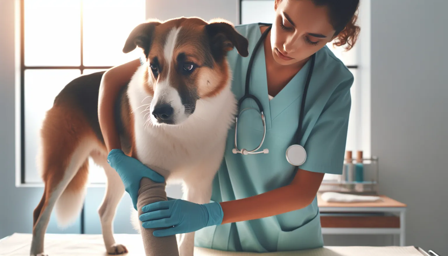
Diagnostic Workup
Orthopedic examination: Elbow pain is assessed by flexion, extension, pronation, and supination of the joint. Pain on full elbow flexion with the wrist supinated is one of the most reliable provocative tests for FCP.
Radiographs: The starting point for most evaluations. Standard elbow radiographs (lateral, cranio-caudal, and oblique views) can reveal secondary arthritic changes, sclerosis of the trochlear notch, and sometimes the fragment itself. However, FCP fragments are often obscured by overlapping bones, making radiographic diagnosis unreliable in early cases.
CT (computed tomography): The gold standard for FCP diagnosis. CT provides cross-sectional imaging that clearly reveals fragments, fissures, and subtle incongruence that radiographs miss. CT changes the surgical plan in approximately 30% of cases compared to radiographs alone.
Arthroscopy: Both diagnostic and therapeutic. Direct visualization of the joint interior reveals the fragment, cartilage damage, and the state of the opposing humeral surface. Many surgeons proceed directly to arthroscopic treatment during the same procedure.
MRI: Useful for detecting cartilage and subchondral bone pathology but less commonly used than CT for FCP due to cost and availability.
Treatment Options
Treatment depends on the severity of fragmentation, the degree of secondary cartilage damage, and the presence of concurrent elbow dysplasia components.
Surgical treatment (recommended for most cases):
- Arthroscopic fragment removal: The most common surgical approach. The loose or fissured fragment is removed, and damaged cartilage is debrided. This is a minimally invasive procedure with rapid recovery.
- Subtotal coronoid ostectomy (SCO): Removal of a larger portion of the coronoid, used when the fragment is large or the remaining coronoid shows widespread pathological changes.
- Proximal ulnar osteotomy (PAUL, BODPUO): A dynamic procedure that alters load distribution across the elbow by cutting the ulna and allowing it to realign. Used when incongruence is a significant contributing factor.
- Sliding humeral osteotomy (SHO): Shifts the load-bearing axis of the humerus to reduce stress on the medial compartment. Considered for advanced medial compartment disease.
Conservative management (when surgery is declined or not appropriate):
- Controlled weight management to minimize joint loading
- Structured, low-impact exercise (swimming, controlled leash walks)
- NSAIDs for pain management (carprofen, meloxicam) under veterinary guidance
- Joint supplements: glucosamine/chondroitin, omega-3 fatty acids, and green-lipped mussel have varying levels of supporting evidence
- Physical rehabilitation: range of motion exercises, therapeutic exercises to maintain muscle mass
Post-surgical reality: Surgery reduces pain and improves function, but it does not prevent the development of secondary arthritis. Virtually all dogs with FCP develop some degree of elbow osteoarthritis over their lifetime. The goal of surgery is to slow the progression and delay the functional impact.
12-Week Post-Surgical Recovery Plan
- Weeks 1-2 (strict rest): Leash walks only for elimination (5-10 minutes, 3-4 times daily). Ice application over the surgical site for 10-15 minutes, 2-3 times daily. Pain management per veterinary prescription.
- Weeks 3-4 (controlled mobilization): Gradually increase leash walk duration to 15-20 minutes. Begin passive range of motion exercises if cleared by your surgeon. No running, jumping, or rough play.
- Weeks 5-6 (gentle strengthening): Introduce short, controlled hill walks. Begin therapeutic exercises (weight shifting, cavaletti poles) if working with a rehabilitation specialist.
- Weeks 7-8 (activity progression): Extend walk duration. Swimming can often begin at this stage (consult your surgeon). Continue building muscle mass around the joint.
- Weeks 9-10 (functional recovery): Most dogs are showing significant improvement by this point. Gradually return to more normal activity levels, avoiding high-impact repetitive exercise.
- Weeks 11-12 (long-term plan): Recheck with surgeon. Establish the long-term exercise, supplement, and weight management plan. Discuss expected arthritis progression and monitoring schedule.
Feeding and Joint Nutritional Support
- Maintain lean body condition throughout life — excess weight is the most modifiable risk factor for elbow arthritis progression
- Avoid overfeeding large-breed puppies; slow, controlled growth reduces elbow dysplasia risk
- Use large-breed puppy formulations with appropriate calcium and phosphorus ratios
For detailed guidance:
- Feeding Guide for Adult Dogs: Maintenance Nutrition Without Drift
- Omega-3 Fish Oil for Dogs: Evidence, Dosing Context, and Safety
- Glucosamine and Chondroitin for Dogs
- Joint Supplements
When to Go to the ER Today
FCP is rarely an emergency, but seek same-day evaluation if:
- Sudden severe lameness with the dog refusing to bear weight on the limb
- Acute swelling of the elbow with heat and severe pain (possible joint infection post-surgery)
- Signs of surgical wound infection: redness, discharge, fever
Related Condition Pathways
- Elbow Dysplasia: FCP is the most common component; other components include ununited anconeal process and osteochondrosis dissecans of the humeral condyle
- Osteochondrosis Dissecans: Frequently concurrent with FCP in the elbow
- Arthritis: The long-term consequence of FCP
- Hip Dysplasia: Often concurrent in the same patient; both are developmental orthopedic diseases of large breeds
Related Breed Longevity Guides
- Labrador Retriever Lifespan & Longevity Guide
- Golden Retriever Lifespan & Longevity Guide
- German Shepherd Lifespan & Longevity Guide
- Rottweiler Lifespan & Longevity Guide
- Bernese Mountain Dog Lifespan & Longevity Guide
- Newfoundland Lifespan & Longevity Guide
Longevity Science Connections
- Joint Screening by Breed: When to X-Ray and How Often
- Elbow Dysplasia in Dogs: Lifetime Load Management
- Canine Physical Rehabilitation: Evidence for Recovery
Frequently Asked Questions
Can FCP be prevented?
Not entirely prevented, because genetics play a major role. However, risk can be reduced by feeding large-breed puppy diets (which control growth rate), maintaining lean body condition, avoiding high-impact exercise during the growth phase, and selecting breeding stock with documented elbow clearances.
Is surgery always necessary?
Surgery is recommended for most FCP cases because the fragment causes ongoing joint inflammation and accelerated cartilage destruction. Conservative management can reduce symptoms but does not address the underlying mechanical problem. Dogs managed conservatively tend to develop more severe arthritis more quickly.
Will my dog be normal after surgery?
Most dogs show significant improvement in comfort and function after surgery. However, “normal” joint mechanics are rarely fully restored, and some degree of arthritis will develop over time. The goal is a comfortable, active dog — not a radiographically perfect joint.
Should I screen my large-breed puppy for elbow dysplasia?
Yes. The Orthopedic Foundation for Animals (OFA) recommends elbow evaluation at 24 months for breeding dogs. Earlier evaluation is appropriate if lameness develops. CT screening is more sensitive than radiographs for detecting early FCP.
Is FCP the same as elbow dysplasia?
FCP is the most common form of elbow dysplasia, but elbow dysplasia also includes ununited anconeal process (UAP) and osteochondrosis of the humeral condyle. A dog can have one or more of these components simultaneously.
Medical Disclaimer
This content is educational and does not replace veterinary diagnosis or treatment. Young large-breed dogs with forelimb lameness need orthopedic evaluation to rule out developmental elbow disease.
References
- Fitzpatrick N et al. Subtotal coronoid ostectomy as management of medial coronoid disease in 263 dogs. Vet Surg. 2009;38(2):233-245.
- Samoy Y et al. Computed tomography findings in 32 dogs with elbow dysplasia. Vet Radiol Ultrasound. 2006;47(3):212-221.
- Burton NJ et al. Comparison of CT arthrography and MRI for detection of canine elbow dysplasia. Vet Rec. 2008;162(22):678-684.
- Temwichitr J et al. Breed-associated risks of common diseases in dogs in the Netherlands. Prev Vet Med. 2010;96(1-2):23-32.
- ACVS (American College of Veterinary Surgeons). Canine elbow dysplasia fact sheet. acvs.org.
Related reads
Related Reading
Continue exploring