Evidence deep dives for Eye Conditions
Pair mechanism-level evidence with practical protocol context before discussing next steps with your veterinarian.
A Red Eye Can Mean Almost Nothing — Or Everything
A red eye can mean almost nothing — or it can mean your dog is hours away from permanent vision loss. That uncertainty is exactly why eye complaints deserve faster triage than most other outpatient issues.
“Eye conditions” covers a wide range of disorders affecting the cornea, lens, retina, eyelids, tear film, and intraocular pressure. Some are mild and treatable with prompt care. Others escalate from discomfort to irreversible damage within a day.
Major categories include:
- Surface disease (conjunctivitis, corneal ulceration, dry eye)
- Pressure disorders (glaucoma)
- Lens disease (cataracts, lens luxation)
- Retinal/optic disease
- Eyelid/conformation disorders
What This Means for the Years Ahead
Eye disease affects far more than vision.
Pain burden can be severe. Corneal ulcers and glaucoma are extremely painful conditions that can reduce appetite, disrupt sleep, and alter behavior in ways that erode daily quality of life.
Vision loss changes function and safety. Dogs that cannot see well bump into things, become anxious, and disengage from activities that keep them physically and mentally healthy.
Many conditions are time-sensitive. Fast treatment often draws the line between preserved vision and permanent loss.
Eye disease may reveal systemic illness. Endocrine, immune, infectious, and age-related diseases can manifest through ocular signs first. The eye exam sometimes catches what the annual physical missed.
Early evaluation protects both sight and overall quality of life.
High-Priority Signs Owners Should Not Ignore
Urgent Warning Signs
- Squinting or keeping eye closed
- Eye rubbing or pawing
- Sudden cloudiness or blue/white haze
- Significant redness
- New light sensitivity
- Thick discharge
Emergency-Level Signs
- Sudden apparent vision loss
- Eye trauma or puncture concern
- Marked globe enlargement
- Severely painful eye with behavior distress
- Fixed dilated pupil with pain
Any painful eye should be treated as urgent same-day. Do not wait for a scheduled appointment.
Common Conditions and Typical Patterns
Conjunctivitis
Inflammation of the conjunctival tissues, presenting with redness and discharge. Can be primary or secondary to allergies, dry eye, irritants, or eyelid problems. Often the least dangerous of the “red eye” causes — but still worth ruling out the more serious ones.
Corneal Ulceration
A defect or injury to the corneal surface. Usually painful.
Signs:
- Squinting
- Tearing
- Blue haze
- Rubbing
Deep or infected ulcers can perforate and threaten the eye within hours. This is not a condition to monitor from home.
Keratoconjunctivitis Sicca (Dry Eye)
Reduced tear production leads to chronic irritation, mucoid discharge, and recurrent surface damage. Most cases require long-term treatment adherence — not a short course of drops and done.
Glaucoma
Elevated intraocular pressure that damages the optic nerve and retina. Often presents with severe pain, red eye, corneal edema, and possible sudden vision loss. Immediate treatment is critical. Hours matter.
Cataracts
Lens opacity that impairs vision progressively or rapidly depending on the cause. Diabetic dogs face higher cataract risk (diabetes).
Eyelid and Conformation Disorders
- Entropion (inward eyelid rolling)
- Ectropion
- Distichia/ectopic cilia
These chronically irritate the cornea and predispose to recurrent disease. Structural problems do not self-resolve.
Breed and Conformation Risk
Certain skull shapes and skin conformations create inherent vulnerability:
- Brachycephalic breeds: more exposure keratitis, higher ulcer risk, tear-film issues
- Small breeds: lens luxation and tear-film disorders in some lines
- Sporting/herding breeds: selected hereditary eye disease risks
Examples:
- French Bulldog: corneal exposure and injury risk
- Cavalier King Charles Spaniel: several ocular disorders overrepresented
- Labrador Retriever: inherited retinal and lens concerns in some populations
Breed risk informs vigilance. It does not replace a direct exam.
Diagnostic Workflow
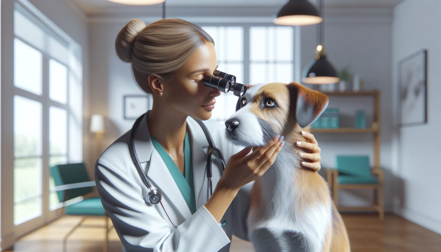
1. Focused Ocular Examination
Covers eyelids, conjunctiva, cornea, anterior chamber, lens, pupil responses, and posterior segment when visible.
2. Essential In-Clinic Tests
Three tests that are fast, inexpensive, and crucial for triage:
- Fluorescein stain (ulcer detection)
- Schirmer tear test (tear production)
- Tonometry (intraocular pressure)
These should be performed on any acutely painful or red eye before treatment decisions are made.
3. Ophthalmic Imaging/Referral (When Needed)
Referral to a veterinary ophthalmologist may be indicated for:
- Suspected glaucoma or lens instability
- Deep or complicated ulcers
- Retinal disease
- Surgical candidates (cataract, eyelid corrections)
Timely specialist involvement can preserve vision in high-risk cases.
Treatment Principles
Treatment depends on the specific diagnosis. One-size-fits-all eye drops can cause harm.
Surface Disease
- Targeted topical antimicrobials when indicated
- Lubrication and tear support
- Anti-inflammatory therapy only when safe for corneal status
Glaucoma/Elevated Pressure
- Immediate pressure-lowering therapy
- Pain control
- Urgent specialist planning for long-term control
Cataracts/Lens Disease
- Monitor functional impact and inflammation
- Specialist evaluation for surgical candidacy where appropriate
Eyelid/Conformation Issues
- Surgical correction may be required for recurrent trauma or chronic irritation
Incorrect medication choice can worsen outcomes. Steroid-containing drops on an active corneal ulcer is the classic example.
The Red-Eye Medication Safety Rule
Never start leftover eye drops in a newly painful eye before a veterinary exam. Steroid-containing drops can rapidly worsen undiagnosed corneal ulceration — turning a treatable problem into a vision-threatening emergency.
For any acute red, squinting, or cloudy eye:
- Prioritize same-day exam with stain, pressure, and tear testing
- Bring all previously prescribed eye medications to the visit
- Restart only medications the current exam confirms are appropriate
This single rule prevents a meaningful share of avoidable vision-threatening complications.
Home Care and Monitoring
Daily Observation
- Redness and discharge trend
- Eye-opening comfort
- Navigation confidence
- Rubbing/squinting frequency
Medication Adherence
- Follow prescribed dosing intervals exactly
- Do not stop early when the eye “looks better”
- Recheck at planned intervals
Environment and Protection
- Reduce trauma exposure in high-risk dogs
- Trim facial hair safely away from the cornea
- Use a harness rather than neck restraint when possible
When Vision Fades: The Adaptation Plan
If vision is reduced, household structure affects long-term quality of life as much as medication adherence.
- Keep furniture layout stable and avoid frequent rearrangement
- Use consistent verbal cues at stairs, thresholds, and feeding areas
- Increase scent- and sound-based enrichment to preserve engagement
Early adaptation reduces secondary anxiety and helps maintain activity as eye disease progresses. Dogs adjust remarkably well when their environment stays predictable.
Prevention and Risk Reduction
True prevention is limited, but early detection is powerful:
- Seek same-day exam for new painful eye signs
- Manage chronic allergy and skin disease that worsens ocular surface inflammation
- Keep endocrine disease controlled (especially diabetes)
- Use routine wellness exams to detect gradual changes early
- Discuss hereditary eye screening in breeding contexts
Prevention here is mostly about risk-aware management and fast response, not total avoidance.
Supplements: Evidence Position
No supplement replaces direct ocular diagnosis and condition-specific treatment. Some nutraceuticals may support general eye health in selected contexts, but high-stakes outcomes in acute eye disease depend on rapid exam and targeted therapy.
Weekly Owner Checklist
- Check for redness, discharge, cloudiness, and squinting.
- Track any bumping into objects or navigation hesitation.
- Keep the medication schedule precise when treating active disease.
- Schedule prompt recheck for any worsening signs.
- Maintain systemic disease control (diabetes, allergies).
Eye Medication Technique That Prevents Treatment Failure
Application quality directly affects outcomes, especially in painful-eye cases:
- wash hands before and after each application
- avoid touching bottle tips to the eye, hair, or skin
- separate multiple drops/ointments by the interval your veterinarian recommends
- store and discard products according to label instructions
Cross-contamination and poor spacing between medications are common reasons “the drops are not working.”
Same-Day Triage Script for Owners
When calling your clinic about an eye problem, give a concise triage summary:
- When signs started and whether they are worsening by the hour
- Pain signs (squinting, pawing, light sensitivity)
- Vision concern (bumping, hesitation, apparent blindness)
- Any prior medications used before the call
Clear communication speeds correct prioritization and reduces delay for vision-threatening disease.
Home Safety Plan for Reduced Vision
If vision is impaired, preserve function and confidence with predictable structure:
- keep walk routes and furniture layout stable
- block access to high-fall-risk zones until navigation stabilizes
- use verbal orientation cues at transitions (doors, stairs, feeding area)
- avoid sudden environmental changes during adaptation weeks
These adjustments reduce secondary injury risk while treatment is underway.
When to Seek Veterinary Care
Urgent Same-Day
- New squinting or eye pain
- Sudden redness or cloudiness
- Thick discharge
- Persistent eye rubbing
Emergency
- Sudden blindness
- Suspected trauma or foreign body penetration
- Severe pain with enlarged eye
- Rapidly worsening eye appearance over hours
With eye disease, delay can mean permanent loss of vision.
What Nutrition Can and Cannot Do
Eye condition management often improves when feeding strategy and medical plan are reviewed together.
- Feeding Guide for Adult Dogs: Maintenance Nutrition Without Drift: is most useful when endpoints are defined before implementation.
- Feeding Guide for Senior Dogs: Healthspan Nutrition: can improve plan adherence when the household needs clear defaults.
- Prescription Diets for Dogs: Evidence, Use Cases, and Limits: supports practical day-to-day decision quality while trend data is gathered.
Confirm timing, dosing, and potential interactions with your veterinarian before adjusting any part of the protocol.
Related Condition Pathways
These condition guides commonly intersect with eye health for diagnostics, prevention, or long-term management:
Related Breed Longevity Guides
These breed longevity guides provide additional context on predisposition patterns and prevention focus:
- French Bulldog
- Cavalier King Charles Spaniel
- Chihuahua
- Golden Retriever
- Labrador Retriever
- Pomeranian
Related Evidence and Research
Frequently Asked Questions
Can eye infections clear without treatment? Some very mild conjunctivitis cases may improve on their own, but this is a risky assumption. A painful, red, or cloudy eye requires a veterinary exam with fluorescein staining and tonometry to rule out corneal ulceration and glaucoma — both of which can look deceptively similar to simple conjunctivitis but progress to permanent vision loss if untreated. The safer approach is always same-day evaluation for any new eye discomfort rather than waiting to see if it resolves.
Is a cloudy eye always cataract? No. Cloudiness in the eye can result from corneal edema (often associated with glaucoma or corneal disease), anterior chamber inflammation, lens sclerosis (a normal aging change), or actual cataract formation. Each cause carries a different prognosis and requires different management. Your veterinarian can distinguish between these with a focused eye exam. Nuclear sclerosis — the most common cause of a bluish haze in older dogs — does not significantly impair vision and does not require treatment.
Can I use leftover eye drops from a prior issue? This is not recommended and can be genuinely dangerous. Steroid-containing eye drops applied to an undiagnosed corneal ulcer can cause rapid worsening, potentially leading to corneal perforation and vision loss within days. Pressure-lowering drops used inappropriately can also cause harm. Every new eye episode should be treated as a fresh problem requiring its own exam and diagnosis before any medication is started.
How fast can glaucoma cause vision loss? Acute glaucoma can cause irreversible retinal and optic nerve damage within hours. It is a true ocular emergency. A dog with a suddenly painful, red, enlarged eye with a fixed dilated pupil needs immediate veterinary care — not a next-day appointment. Even with aggressive treatment, vision cannot always be saved once intraocular pressure has been critically elevated, which is why speed of response is the primary determinant of outcome.
Do all cataracts require surgery? Not all cataracts need surgical removal. The decision depends on how much the cataract impairs vision, whether it is causing secondary inflammation inside the eye, the dog’s overall health and anesthetic candidacy, and the ophthalmologist’s assessment of surgical benefit versus risk. Many dogs with partial or slowly progressing cataracts maintain functional vision with monitoring alone. Dogs that are good surgical candidates and have significant vision impairment often regain excellent visual function after phacoemulsification performed by a veterinary ophthalmologist.
When should I see a veterinary ophthalmologist? Referral to a veterinary ophthalmologist is warranted for suspected glaucoma, deep or complicated corneal ulcers, lens luxation, retinal disease, cases requiring cataract surgery evaluation, and any eye condition that is not responding as expected to initial treatment. Ophthalmologists have specialized equipment and training that allows more precise diagnosis and access to surgical options not available in general practice. Timely specialist involvement often draws the line between preserved and lost vision in high-stakes cases.
Medical Disclaimer
This guide is informational and does not replace in-person veterinary diagnosis or treatment. If your dog is acutely unwell, seek veterinary care immediately.
References
[1] Merck Veterinary Manual: Eye Disorders in Dogs [2] American College of Veterinary Ophthalmologists (ACVO) [3] AAHA Canine Life Stage Guidelines [4] WSAVA Global Nutrition Guidelines [5] American College of Veterinary Internal Medicine (ACVIM) [6] Dog Aging Project
Related reads
Related Reading
Continue exploring